
Research Signal – Source date 2022-11-05
Age-related macular degeneration (AMD) damages the macula, the central part of the retina responsible for detailed vision. In its early stages, deposits called drusen can form around the retinal pigment epithelium, or RPE. Later disease can involve either gradual loss of photoreceptors in dry AMD or abnormal blood-vessel growth in wet AMD. Both forms can threaten central sight.
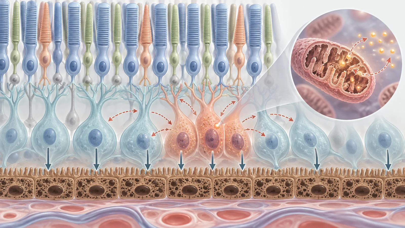
The RPE is a layer of support cells beneath the light-sensing photoreceptors. It helps maintain the blood–retina barrier, clears away the used outer segments of photoreceptors, and participates in the visual cycle that prepares light-sensitive molecules for continued vision. When RPE function declines, photoreceptors lose part of the environment that keeps them working.
A review published on November 5, 2022, brings together evidence that connects oxidative stress with a cellular state called senescence in the aging retina. Senescent cells have stopped dividing, but they are not simply dead cells. They remain metabolically active and can change their surroundings by releasing inflammatory signals. The idea raises an important treatment question: could some living but dysfunctional retinal cells become targets before they contribute to further damage?
Why the aging retina becomes vulnerable
Oxidative stress occurs when the production of reactive oxygen species, or ROS, outweighs the body’s antioxidant defenses. ROS are chemically reactive molecules produced in part by mitochondria, the structures that help cells make energy. Aging can weaken antioxidant protection, leaving retinal cells more exposed to molecular damage.
The relationship can become self-reinforcing. ROS can damage mitochondrial DNA and other mitochondrial components. Damaged mitochondria may then function poorly and generate still more ROS. The review links this feedback loop with inflammatory signaling involving pathways such as NF-κB, the NLRP3 inflammasome, and cGAS/STING. These pathways can alter how cells respond to stress and can help sustain inflammation.
Oxidative stress also affects the systems that dispose of damaged material. In RPE cells, insufficient autophagy can allow ubiquitinated proteins and other cellular debris to accumulate. The review connects this impaired cleanup with drusen formation and RPE dysfunction. These processes are not separate from the wider biology of aging: damaged proteins, impaired mitochondria, and inflammatory signals can all make it harder for retinal support cells to maintain their normal duties.
How a stressed cell can keep affecting its neighbors
Cellular senescence is a response to stress characterized by cell-cycle arrest. The review distinguishes stress-induced premature senescence from replicative senescence, which is associated with repeated cell division and telomere shortening, and from developmentally programmed senescence. Oxidative stress is presented as a trigger for the stress-induced form.
A senescent cell can release a collection of inflammatory cytokines, growth factors, and enzymes known as the senescence-associated secretory phenotype, or SASP. Reported SASP components include IL-6, IL-8, transforming growth factor beta, matrix metalloproteinases, and vascular endothelial growth factor. These signals can recruit immune cells, intensify inflammation, and influence nearby cells. In some experimental settings, SASP from senescent cells can push otherwise healthy cells toward senescence-associated changes as well.
This makes senescence different from a single episode of injury. A damaged cell that dies is no longer an active source of signaling. A senescent cell may remain in the tissue, stop dividing, and continue altering its local environment. The proposed concern in the retina is therefore not only whether oxidative stress injures RPE cells, but whether surviving stressed cells can maintain conditions that undermine neighboring cells and retinal function.
What the retinal evidence shows
The review brings together several kinds of observation, and they answer different questions. In primates, senescent RPE cells were observed around drusen. In people with geographic atrophy, RPE tissue showed increased expression of the senescence marker p16 compared with age-matched controls. Those findings place senescence-associated features near retinal changes relevant to AMD.
Experiments in cultured RPE cells show how stress can produce similar features under controlled conditions. Hydrogen peroxide and cigarette smoke increased markers including SA-β-Gal, p16, p21, γH2AX, and SASP-related signals in ARPE-19 cells. Hydrogen peroxide was also linked with activation of the p53/p21 pathway. RPE cells that ingested oxidized photoreceptor outer segments developed stress-induced senescence-associated changes and altered secretion of factors including TNF-α, IL-8, VEGF, and complement factor H.
These observations support a biologically plausible connection between oxidative stress and senescence in retinal support cells. They do not, by themselves, establish the complete sequence in patients. A higher marker level in tissue indicates a cellular state or response; it does not prove that the marked cells initiated AMD, that they are sufficient to drive progression, or that removing them would preserve a person’s vision. The evidence spans human tissue, primates, cultured cells, and animal models, so the results should be read according to what each setting can actually show.
Photoreceptors also require careful interpretation. The review describes an experiment in an immortalized mouse cone-cell line in which an oxidative-stress-inducing compound led to apoptosis through increased p53 activity. That result shows that oxidative stress can contribute to photoreceptor cell death in an experimental system. It does not establish that the photoreceptors underwent senescence before dying.
Changing cell behavior is not the same as removing cells
Potential interventions fall into more than one category. Some aim to reduce the conditions that sustain senescence or to suppress the inflammatory behavior of senescent cells. Others, called senolytic strategies, aim to eliminate senescent cells selectively.
The review describes rapamycin as an example of the first approach in RPE cells. Rapamycin inhibited mTORC1 signaling, reduced SA-β-Gal activity and p16 expression, and activated autophagy in the experimental setting discussed. The reported findings suggest that changing nutrient-response and cleanup pathways can alter senescence-associated behavior. They do not establish a treatment regimen for people with AMD.
Senolysis presents a different challenge. Genetic systems such as INK-ATTAC and p16-3MR, along with drugs aimed at survival pathways such as the BCL-2 family, have been used to remove senescent cells in experimental models. The attraction is clear: if a harmful cell population is actively maintaining inflammation, clearance might reduce that influence.
Selectivity is the central difficulty. Senescence also has physiological roles, including wound repair, tissue remodeling, and tumor suppression. A therapy would need to distinguish disease-driving senescent cells from cells participating in useful biological responses. The review describes the therapeutic concept and experimental rationale, but the supplied evidence does not establish that broad removal of senescent cells is safe or beneficial in human AMD.
What the vascular model can and cannot tell us
One of the clearest clearance results discussed in the review comes from oxygen-induced retinopathy, an animal model considered in relation to proliferative diabetic retinopathy. In that model, senescence markers and SASP-related signals increased in several cells that form retinal vascular units, including endothelial cells, pericytes, astrocytes, and Müller glia. Removing senescent cells with genetic or drug-mediated approaches suppressed the pathological vascular response and supported healthier reparative vascular remodeling in mice.
That result gives senolysis a vascular-remodeling rationale in a particular experimental disease model. It is not a direct demonstration that senescent-cell clearance treats AMD. Oxygen-induced retinopathy, proliferative diabetic retinopathy, and AMD involve different disease contexts and endpoints. A change in abnormal vessels in mice is also not the same as demonstrated preservation of central vision in patients.
The review also records a phase 2 trial of an intravitreal BCL-xL inhibitor in people with neovascular AMD as ongoing at the time of publication. This places senescent-cell targeting within clinical investigation, but it is not evidence of demonstrated safety, efficacy, or improved vision. The authors conclude that the mechanisms linking oxidative stress-induced senescence to AMD remain unresolved and require further work before the translational implications can be settled.
What targeting senescent cells could mean for preserving sight
The practical promise is preservation rather than restoration. If researchers can identify which stressed retinal cells are actively driving harmful inflammation, a future treatment might reduce those cells or quiet their damaging signals before more central retinal function is lost. That could matter for people at risk of further vision loss from disease affecting the macula.
The possibility remains conditional on selective and safe targeting, and on clinical studies showing a meaningful visual benefit. The 2022 review supplies a connected biological hypothesis and several experimental clues, not a validated AMD therapy. It points toward a treatment strategy that would protect remaining retinal function only if the relevant disease-driving cells can be distinguished from useful senescent responses and the intervention proves effective in patients.
Key Sources
- Primary source: Oxidative Stress-Induced Cellular Senescence in Aging Retina and Age-Related Macular Degeneration.
DOI or Journal Page · Review or Meta-Analysis · 2022-11-05
Keep exploring