
Research Signal – Source date 2025-09-12
Osteoarthritis is often described as a condition in which the cushioning cartilage in a joint wears down. Extra body weight can increase the force a joint carries, making mechanical loading an important part of the explanation. But the September 2025 review The Bidirectional Role of Obesity and Aging in the Pathogenesis of Osteoarthritis examines another possibility: fat tissue may also send inflammatory signals that affect cartilage, while aging-related changes may feed back into metabolic inflammation.
The authors describe a structured search of PubMed, Embase, and Web of Science and selected peer-reviewed articles published from 2013 through 2023. Their synthesis brings together different kinds of evidence, including mechanistic, animal, cell, observational, and cited clinical treatment studies. It offers a way to connect obesity, aging, and joint damage, while leaving important questions about cause and treatment unresolved.
Fat tissue can send signals beyond the joint
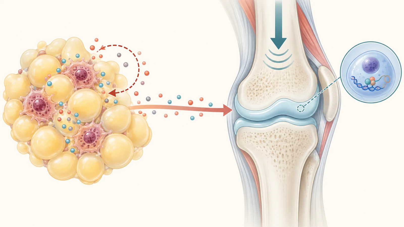
Adipose tissue—the body’s fat tissue—is more than an energy store. It contains fat cells alongside immune and nerve cells, and it can release signaling molecules into the body. The review describes obesity as a state of low-grade, chronic inflammation in which this tissue’s balance of immune activity and secreted molecules can change.
One change discussed is a shift in the mix of macrophages, immune cells found in adipose tissue. In lean tissue, alternatively activated M2 macrophages are associated with anti-inflammatory signals. In obesity, the balance can move toward classically activated M1 macrophages, which release inflammatory molecules such as interleukin-1 (IL-1), IL-6, and tumor necrosis factor alpha (TNF-α). These signals can help sustain inflammation beyond the fat tissue itself.
Fat cells also release adipokines, signaling molecules that include leptin and adiponectin. In the pathway summarized by the review, inflammatory cytokines can activate signaling inside joint tissues, including p38 and ERK1/2 pathways. This activity is associated with increased matrix metalloproteinases (MMPs), enzymes that break down components of cartilage’s structural matrix.
Leptin is one example the authors discuss in more detail. They report that levels are elevated in people with osteoarthritis compared with healthy individuals and that leptin is present in joint cartilage and the synovium, the tissue lining the joint. The review describes findings linking leptin to increased activity of cartilage-degrading enzymes and loss of proteoglycans, molecules that help cartilage retain its structure. These findings provide a plausible biological route from obesity-related signals to cartilage breakdown, alongside the added physical load on a joint.
Aging may feed inflammation back into metabolism
The review’s proposed return path begins with cellular senescence. Senescent cells have stopped dividing, but they can remain active and release a mix of inflammatory cytokines and other bioactive molecules, often called the senescence-associated secretory phenotype, or SASP. Senescence has normal roles in processes such as development and tumor suppression; the concern is that senescent cells may accumulate with age and contribute to chronic disease.
In adipose tissue, the review links a higher burden of senescent cells with local and systemic inflammation. It also describes aged fat-cell progenitors—cells that can develop into fat cells—as having reduced capacity to differentiate. Signals they release, including activins, may affect nearby cells’ insulin sensitivity and attract immune cells. The proposed result is worsening metabolic dysfunction and insulin resistance, which can add to the inflammatory environment associated with obesity.
This aging-to-fat-tissue route is related to, but distinct from, the route from fat tissue toward cartilage. The review presents senescent cells and their secretions as a possible way aging could amplify obesity-associated inflammation and metabolic problems. It does not show that the complete cycle—obesity-related signals damaging joints, followed by aging-related changes reinforcing those signals—has been demonstrated in people with osteoarthritis.
Gene regulation may shape cartilage’s response
Inflammatory and metabolic stress can matter to cartilage partly through changes in how cells regulate genes. Epigenetic regulation affects whether genes are more or less active without changing the underlying DNA sequence. The review discusses DNA methylation, a chemical modification that can influence gene activity, as one mechanism associated with cartilage breakdown.
In healthy cartilage, the genes for many matrix-degrading enzymes are kept relatively quiet. The review describes osteoarthritis-related findings in which higher levels of MMP-3, MMP-9, and ADAMTS-4 are associated with demethylation of gene promoters. Promoters act like control regions for gene activity. If their regulation changes, a chondrocyte—the cell that maintains cartilage—may produce more of the enzymes involved in breaking down the surrounding matrix.
Non-coding RNAs offer another layer of control. These RNA molecules do not provide instructions for making proteins, but they can help regulate gene expression. The review names miR-140 and miR-146a among non-coding RNAs whose regulation is altered in osteoarthritis. Such changes may be part of how cartilage cells respond to aging, obesity, or inflammation, though the precise links remain under study.
The authors emphasize that associations between epigenetic markers and disease do not settle which changes drive cartilage damage and which may follow it. Epigenetic regulation can be reversible, making it an area of therapeutic interest, but changing a marker has not been shown here to prevent or reverse osteoarthritis damage in people.
Treatment ideas are at different stages
Current osteoarthritis care includes approaches aimed at symptoms, along with injections and emerging molecular strategies. The review notes that several experimental disease-modifying approaches have shown potential in preclinical or early clinical research, but none of the compounds it discusses has received regulatory approval for clinical use as a disease-modifying osteoarthritis drug. The proposed obesity–aging mechanisms therefore remain some distance from an established treatment that changes the course of joint damage.
The human injection studies cited in the review do not point to one consistent result. One study of 95 people with mild to moderate osteoarthritis compared corticosteroids, hyaluronic acid, platelet-rich plasma, and placebo; the review reports no significant changes in knee outcomes in either the short or long term. Other cited studies report improvements after injections, including in pain, stiffness, or function. These varied findings concern different treatments and groups of patients. They do not show that an injection acts on the obesity–aging pathway or slows cartilage degeneration through that mechanism.
Senescent-cell approaches are at an earlier experimental stage for this question. The review describes animal research in which removing senescent cells improved metabolic measures in obese models, as well as cell studies in which dasatinib and quercetin reduced senescent-cell burden and inflammatory markers in human adipose tissue cells. These results are not evidence that clearing senescent cells benefits people with osteoarthritis. Animal models also do not fully reproduce human cartilage structure and responses, a limitation the authors say supports developing more representative models.
The proposed chain still needs a causal test
The review organizes evidence from studies that differ in design, population, and model. Much of the epidemiological evidence it discusses is cross-sectional: it captures associations at a point in time, rather than showing how obesity, aging, and joint damage change in sequence. As a result, it cannot by itself establish which factor came first or whether one caused another. The authors also note that associations may differ across populations.
The molecular findings help identify plausible links: inflammatory signals from fat tissue can relate to cartilage-degrading activity, while senescent cells may contribute to inflammation and metabolic dysfunction. But evidence for separate links does not confirm the complete two-way process in people. Likewise, altered DNA methylation or non-coding RNA activity alongside osteoarthritis does not establish that those changes cause damage or that targeting them would change disease outcomes.
Longitudinal studies that follow people over time could help clarify how these factors develop together. The review also calls for larger, more diverse research and for better ways to connect molecular findings with clinical outcomes. Its synthesis makes the biological case for looking beyond joint load alone, but the relative contribution of each route—and whether interrupting one can protect a human joint—remains unsettled.
Could treatment slow osteoarthritis damage?
For people with osteoarthritis, the hoped-for benefit is care that can do more than ease symptoms: it might also protect cartilage by addressing the inflammatory and age-related processes affecting a particular joint. That possibility depends on identifying which pathways truly drive damage in people and then showing that interventions aimed at them are safe and effective. The review brings those research directions into view, but does not yet establish that such protection is within clinical reach.
Key Sources
- Primary source: The Bidirectional Role of Obesity and Aging in the Pathogenesis of Osteoarthritis: Molecular Mechanisms, Epigenetic Insights, and Therapeutic Implications.
DOI or Journal Page · Review or Meta-Analysis · 2025-09-12
Keep exploring