
Research Signal – Source date 2022-03-15
Some people naturally sleep fewer than six and a half hours a night without an obvious cost to their cognition. Researchers have identified mutations associated with this familial natural short sleep, or FNSS, and have used them to ask a more specific question: can the biology associated with needing less sleep also alter the brain changes associated with Alzheimer’s disease?
A mouse study published on March 15, 2022, explored that question with two FNSS mutations. The result was a reduction in several Alzheimer-like pathology measures, including tau deposits and amyloid plaques. But the experiment tested inherited mutations in carefully constructed mouse models. It did not test whether deliberately sleeping less protects the human brain, or whether the mutations worked by making sleep more restorative.
A genetic experiment, not a sleep-restriction test
The researchers began with two mouse lines carrying different FNSS mutations. One changed the DEC2 gene, producing the DEC2-P384R variant. The other changed Npsr1, producing Npsr1-Y206H. These genes were used as biological models of inherited short sleep, not as a prescription for reducing sleep time.
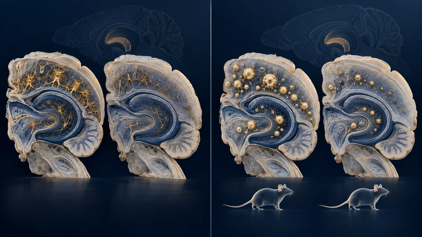
Each mutation was then crossed separately with mouse models designed to reproduce different components of Alzheimer-like pathology. The PS19 model develops abnormal tau accumulation. The 5XFAD model develops amyloid-beta, or Aβ, plaques. Using both models allowed the researchers to examine whether the FNSS mutations affected tau and amyloid-related changes in similar or different ways.
The comparisons were made against control genotypes within the same experimental systems. This matters because a genetic background can influence pathology independently of sleep. The study therefore provides evidence about interactions between particular mutations and particular mouse disease models, rather than a general result about all people who sleep for fewer hours.
Tau and amyloid did not follow one identical pattern
At six months, both FNSS mutations were associated with less tau pathology in the hippocampus of PS19 mice. For DEC2-P384R, the researchers found significantly less phosphorylated tau deposition in the dentate gyrus than in the corresponding DEC2-WT;PS19 controls. Npsr1-Y206H;PS19 mice also showed diminished tau phosphorylation in the same region compared with PS19 controls.
The amyloid findings were more conditional. At three months, there was little plaque deposition across the DEC2-related 5XFAD groups. By six months, plaques were clearly present, and DEC2-P384R;5XFAD mice had less plaque-covered area in both the cortex and hippocampus than DEC2-WT;5XFAD mice. The comparison was specifically with the DEC2 wild-type control, not simply with an untreated or unrelated group.
The second mutation did not produce an identical result across sexes. Female Npsr1-Y206H;5XFAD mice had significantly less plaque coverage in the hippocampus or cortex than 5XFAD controls. The male comparison did not reach statistical significance. Female 5XFAD mice develop more robust pathology than males in this experimental context, but statistical significance in females and its absence in males do not by themselves establish that the mutation’s effect differs between sexes.
There was also an interpretive wrinkle in the DEC2 experiment. Plaque formation was higher in DEC2-WT;5XFAD mice than in 5XFAD mice without the added DEC2 transgene. That observation suggests that wild-type DEC2 may itself influence the amyloid-aggregation pathway. It does not erase the lower plaque load seen with DEC2-P384R, but it makes the relevant comparison the mutant-versus-matched-wild-type result rather than a simple ranking of every mouse line.
The protein measurements changed with age and compartment
Visible deposits were only one part of the study. The researchers also measured different forms of tau and Aβ42 in brain tissue and cerebrospinal fluid, and those measurements did not always move in the same direction as plaque or tangle staining.
In the DEC2-P384R;PS19 experiment, three-month-old mutant and control mice had similar amounts of total tau across the tested fractions, indicating that tau expression from the transgene was maintained. Yet the mutant mice had more phosphorylated tau in the soluble fraction. By six months, when pathology was more established, the pattern had shifted: DEC2-P384R;PS19 mice had less total tau in the insoluble fraction and less phospho-tau in both the less-soluble and insoluble fractions, with lower phospho-tau-to-total-tau ratios in both fractions. The important difference was therefore not simply “more protein” or “less protein,” but where the modified tau was found and how much had accumulated in harder-to-solubilize forms.
Npsr1-Y206H did not mirror every DEC2 result. At three months, Npsr1-Y206H;PS19 mice had lower phospho-tau in less-soluble and insoluble fractions. At six months, the significant difference was confined to the insoluble fraction. These age-specific measurements show why a single time point cannot stand in for the entire disease process, and why a lower deposit burden does not require every related protein concentration to be lower at every stage.
The DEC2 amyloid measurements were similarly time-sensitive. At three months, DEC2-P384R;5XFAD mice had higher Aβ42 in extracts from the cortex and hippocampus and more Aβ42 in cerebrospinal fluid than their DEC2-WT;5XFAD controls. Those differences were no longer present in the same form at six months, when plaque load was lower in the mutant mice and tissue Aβ42 did not differ between the relevant groups. Npsr1-Y206H;5XFAD mice instead showed higher cerebrospinal-fluid Aβ42 at six months. Concentration, location, age and visible accumulation were related measurements, not interchangeable ones.
The surrounding tissue also showed signs of less damage
The Npsr1-Y206H;PS19 mice offered evidence beyond counting tau deposits. In six-month-old PS19 mice, staining for synaptophysin was reduced in the CA3 region, consistent with loss or deterioration of synaptic structures. The signal was not reduced in the Npsr1 mutant PS19 mice. Synaptophysin is a marker associated with nerve-terminal structures, so the result suggests that the mutation was accompanied by preservation of a tissue feature affected in the PS19 model.
The researchers also measured GFAP, a marker used here to assess astrogliosis, the response of astrocyte cells to disturbance or injury. GFAP signal was lower in the hippocampus of Npsr1-Y206H;PS19 mice than in PS19 mice, and the difference was also supported by Western blotting. Together, the synaptophysin and GFAP findings are consistent with less synaptic loss and astrogliosis in this particular double-mutant model.
They remain tissue measurements. They do not demonstrate improved memory, preserved learning, or a cognitive benefit in the mice. The study’s pathology results and its behavioral conclusions should therefore be kept separate.
Fluid levels left the clearance story open
A concentration in cerebrospinal fluid is not a direct measurement of how quickly a protein is produced, transported, degraded or cleared. The study’s fluid results illustrate why the mechanism remained unsettled.
For DEC2-P384R;5XFAD mice, higher brain and cerebrospinal-fluid Aβ42 at three months preceded the lower plaque burden measured at six months. In Npsr1-Y206H;5XFAD mice, cerebrospinal-fluid Aβ42 was higher at six months while plaques were reduced in the female comparison. Those patterns could be compatible with altered movement or disposal of Aβ42, but they do not identify a clearance rate or establish one shared process between the two mutations.
The tau experiments made the same point in another way. Npsr1-Y206H;PS19 mice had lower cerebrospinal-fluid tau than control mice. In isolated hippocampal nerve terminals, baseline human tau release was similar between groups, but treatment with the Npsr1 ligand NPS increased tau release only in the mutant synaptosomes. Greater stimulated release at the nerve terminal therefore coexisted with lower cerebrospinal-fluid tau in the animals.
The authors raised alternative explanations, including the possibility that extracellular tau was being degraded within glial cells or handled through another pathway in the mutant brain. The measurements narrow the possibilities, but they do not prove that the two mutations improve clearance in the same way.
The missing link is whether sleep biology caused the protection
The central unresolved question is what the mutations changed. They may alter pathways connected with sleep, neuronal activity, protein handling or other functions. The experiment did not isolate sleep duration or demonstrate that enhanced restoration during sleep produced the lower pathology measures. Because the mutations were inherited and present throughout the animals’ development, the findings cannot be treated as a test of voluntary sleep restriction.
The authors specifically called for detailed sleep studies using EEG and EMG, along with cognitive and behavioral analysis of the double-mutant mice. They also identified the need for additional FNSS and Alzheimer-like models, and for experiments that can control when and where the relevant mutations are active. Those approaches would help separate an effect of sleep-related physiology from effects that arise elsewhere in the nervous system.
The evidence came from several assays with different sample sizes. For example, the six-month DEC2-P384R;PS19 tau measurements used 10 mice per group, while the three-month groups used four mice per group in the reported biochemical analyses. The plaque comparisons included different numbers of animals and brain slices across genotypes and sexes. The immunohistological methods separately reported that data analysis was blinded to genotype. These details do not turn the study into one uniform experiment with one universal statistical strength. They show why each result needs to remain tied to its model, age, sex and assay.
Nor did the human family observations answer the epidemiological question. The authors noted that their database of FNSS families was not large enough to test whether these individuals have lower dementia risk. The mouse results were therefore a starting point for investigating protective biology, not evidence that natural short sleepers are protected from Alzheimer’s disease.
What protective sleep biology could change
The practical promise of this line of research is the possibility of preserving brain function by identifying biological processes associated with restorative sleep and resistance to tau or amyloid pathology. People at risk of neurodegeneration could eventually benefit if researchers can determine which pathways are responsible, establish whether sleep actually mediates their effects, and safely influence those pathways in humans with meaningful benefit.
In March 2022, that possibility remained conditional. The mutations had changed pathology in specific mouse models, but the responsible mechanisms, their relationship to sleep, and their relevance to human treatment still needed to be established.
Key Sources
- Primary source: Familial natural short sleep mutations reduce Alzheimer pathology in mice.
DOI or Journal Page · Peer-Reviewed Paper · 2022-03-15
Keep exploring