
Research Signal – Source date 2021-02-01
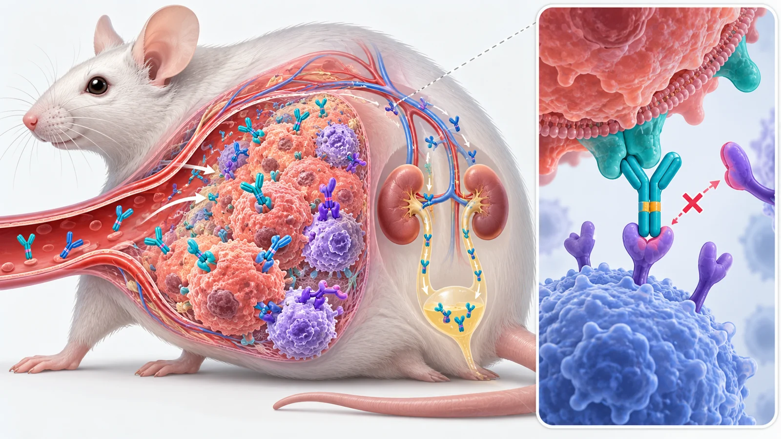
A labeled nanobody identified tumors expressing PD-L1 in mice as soon as one hour after injection. In the reported comparison, it detected PD-L1-positive tumors but not tumors in mice lacking PD-L1, supporting the tracer’s target specificity. The result is striking, but it answers an imaging question: where is this immune checkpoint found? It does not show that target detection produces a treatment benefit.
The finding appears in a February 2021 review of research on nanobodies aimed at immune checkpoints. The studies it brings together range from binding tests and cell experiments to mouse tumor treatment and imaging. They explore two related possibilities, but they do not provide one clinical test of both.
An immune brake becomes a target
Immune checkpoint proteins help regulate immune activity. Some transmit inhibitory signals when they bind a partner molecule, restraining immune cells. Tumors can make use of these brakes to limit an antitumor response. Checkpoint-blocking drugs aim to interrupt that restraint so immune cells can act against cancer.
The review focuses on nanobodies, small antibody fragments built from a single antigen-binding domain. Conventional antibodies have more parts; a nanobody can still recognize a particular target while weighing about 15 kilodaltons, roughly one-tenth as much as a conventional antibody. That size matters in different ways for imaging and treatment.
For a scan, a nanobody can be attached to a detectable label. It travels into tissue and binds its target. The review links its small size with deeper tumor penetration, while unbound nanobodies are rapidly cleared, mainly through the kidneys. Less lingering tracer away from the target can improve the contrast between a tumor signal and background. These properties help explain why a scan can show a target quickly; they do not show that binding changes the tumor’s course.
Mouse scans show where checkpoints are
In the PD-L1 study summarized by the review, researchers attached a radioactive label to selected nanobodies and used SPECT/CT scans to see where the probes accumulated in mice. The C3 and E2 tracers showed useful target binding and distribution. In a mouse tumor model, the labeled probes detected PD-L1-expressing tumors, while the knockout comparison provided a specificity check. The review reports that imaging could reveal the target as soon as an hour after injection.
A separate set of experiments looked at LAG-3, another checkpoint molecule. Nanobodies raised against mouse LAG-3 were labeled for SPECT/CT. In mice, the researchers observed tracer uptake in immune organs and in a subcutaneous tumor engineered to overexpress LAG-3. The review identifies nanobodies 3132 and 3206 as effective tools for evaluating LAG-3 expression in that setting, with imaging contrast visible at an early time point.
These experiments used different targets and models. Both show that labeled nanobodies can locate checkpoint expression in mice; neither measures whether a treatment is working. A tracer’s presence says that it reached and bound a target. It does not say whether blocking that target will activate a useful immune response, or whether a scan can guide care in people.
The review also describes a fluorescent nanobody complex that detected CTLA-4-positive T cells in cell-based tests. That work demonstrates another way to see target-bearing cells, but it did not provide real-time tracking of changing checkpoint levels in a living tumor. Imaging a target in a sample and following its changes throughout treatment are different capabilities.
Treatment asks more than binding
A therapeutic nanobody must do more than find a checkpoint. It must interfere with the relevant signal in a way that helps antitumor immunity, and its effects have to last long enough to matter. Results from treatment experiments therefore address a different question from tracer studies.
The review describes four CTLA-4-binding nanobodies from a camel immune library. One, Nb16, reduced tumor growth and prolonged survival in melanoma-bearing mice. In the comparison reported, however, the nanobody and monoclonal-antibody groups both showed antitumor effects, with no difference between them. The experiment is evidence of activity in mice, not evidence that the smaller format worked better than the conventional antibody.
Other examples illustrate how incomplete the path from target recognition to treatment can be. In one lab study, an anti-TIM-3 nanobody slowed the growth of a leukemia cell line. In a separate study of nanobodies against the same checkpoint, researchers measured binding but did not test whether the molecules could activate antitumor T cells. The review also describes PD-L1 nanobodies whose binding was measured without testing whether they blocked checkpoint signaling. Binding alone cannot establish a treatment effect.
Fast clearance helps scans and complicates treatment
A tracer benefits from clearing quickly: once unbound probe leaves the body, the target-bound signal can stand out sooner. For treatment, brief exposure may be less useful if sustained checkpoint blockade is needed. The same rapid clearance that helps produce an early image can therefore create a design challenge for a drug.
The studies summarized in the review describe ways to extend nanobody exposure. One anti-PD-1 nanobody, Nb97, was combined with human serum albumin in a fusion protein; the review reports that this extended its serum half-life. The review also describes nanobody dimers and multimers as approaches to increasing molecular size and prolonging time in the body. These are design strategies, not established clinical solutions. Changing size can affect how a molecule distributes and clears, so imaging advantages and treatment requirements need not point to the same format.
Repeated measurement remains a clinical aim
Checkpoint expression can vary between tumors and change over time, making repeated measurement appealing. A scan that could reliably show those changes might help clinicians assess whether a tumor carries a target before treatment or how target expression shifts during therapy. A tissue sample offers a limited view; an imaging method could potentially examine target distribution across the body.
The 2021 review describes early mouse imaging snapshots and cell-based detection, not validated repeated scans in patients. A one-hour scan establishes neither accuracy over time nor usefulness for choosing or adjusting treatment. Those would require reliable imaging in people and evidence that the information improves a meaningful clinical decision.
What quicker checkpoint scans could change
If nanobody imaging can reliably show checkpoint expression throughout a person’s tumors, clinicians could have a noninvasive way to assess targets before or during immunotherapy. That could help connect treatment choices to where an immune brake is present and whether its distribution changes. The mouse scans in the review make this a plausible research direction, while human reliability and clinical value remain the conditions for such use. This is the perspective of the February 2021 review, not a claim of later progress.
Key Sources
- Primary source: Nanobodies targeting immune checkpoint molecules for tumor immunotherapy and immunoimaging (Review).
DOI or Journal Page · Review or Meta-Analysis · 2021-02-01
Keep exploring