
Research Signal – Source date 2024-12-26
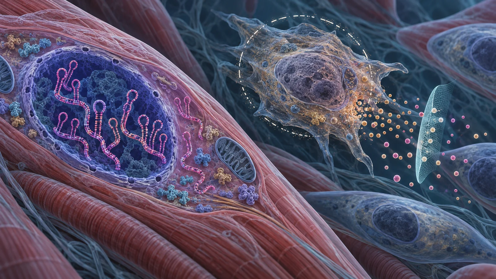
Myotonic dystrophy type 1 (DM1) begins with an inherited genetic change, yet its effects can reach far beyond muscle. People may experience muscle weakness and stiffness, alongside problems involving the heart, breathing, digestion, vision, cognition, or metabolism. The mix and severity vary widely. A review published on December 26, 2024, considers one possible contributor to that range: cells that show features of premature aging and release inflammatory signals.
The idea is that toxic RNA produced by the altered gene may push some cells into a lasting stop in cell division, called cellular senescence. Such cells can also release a blend of signaling molecules known as the senescence-associated secretory phenotype, or SASP. If this process contributes to damage in more than one tissue, treatments that remove senescent cells or quiet their signals might eventually complement approaches aimed at the toxic RNA itself.
That is a therapeutic hypothesis, not a result in people with DM1. The evidence brought together in the review includes observations in patient-derived cells and tissue, laboratory models, and a fly experiment. It does not show that a senescence-targeting treatment improves DM1 symptoms in people.
One inherited repeat can have many effects
DM1 is caused by an expansion of CTG repeats in the DMPK gene. The expanded gene produces RNA containing repeated CUG sequences. This RNA accumulates inside cells and can trap proteins that normally help process other RNA messages. With those regulators disrupted, many genes are spliced incorrectly, changing how their instructions are assembled. The review describes additional effects on gene expression and RNA handling, all part of a broader cascade of RNA toxicity.
The expansion is inherited, but the illness does not follow one simple path from repeat length to symptoms. DM1 has different forms based on age at onset, and manifestations and progression vary. The review notes that repeat length has only a moderate relationship with clinical features. A single genetic cause therefore leaves open questions about why particular organs are affected and why the burden differs so much among people.
Cellular senescence is one proposed piece of that puzzle. Senescent cells have stopped dividing, often in response to stress, and can remain metabolically active. Their SASP can include inflammatory cytokines, chemokines, and other molecules. Those secretions may influence nearby cells and the tissue environment, making senescence potentially relevant beyond the cell that first changes. But finding senescence-associated features does not by itself tell researchers how much this process contributes to DM1’s symptoms.
Patient-derived cells show a slower-growing pattern
Across studies summarized in the review, myoblasts taken from skeletal muscle of people with DM1 grew more slowly than comparison cells. The observations included cells from congenital DM1 and from several muscles in people with DM1. Primary fibroblasts from patients also showed reduced growth. In some samples, cells appeared larger and flatter, a shape commonly associated with senescence.
Other cellular features point in the same direction. DM1 fibroblasts showed changes in nuclear shape and in the location of proteins that help form the nuclear envelope. Investigators also reported increased senescence-associated beta-galactosidase activity in patient-derived muscle cells and fibroblasts, along with shifts in cell-cycle regulators. These included higher levels of inhibitors such as p16 and p21 in several examined materials. In muscle sections, a larger proportion of satellite cells, which help repair muscle, expressed p16.
Inflammatory signals add another layer. Studies of DM1 myoblasts found increased expression of molecules including CXCL1, CXCL8, IL-6, and MMP1. Other patient-derived cells and models showed changes in IL-6 and additional inflammatory factors. In one adult cohort, higher IL-6 in blood was associated with lower muscle strength and physical function. An association can make a process worth investigating, but it cannot show that IL-6 or senescence caused the functional differences.
These results come from different tissues, cell types, and experimental settings. Their value is that several kinds of observation—slow growth, altered cell-cycle markers, and inflammatory output—fit a senescence-related pattern. They do not establish that every affected organ contains the same cells or that the pattern accounts for the full range of disease.
A proposed route from RNA stress to inflammation
The review outlines a possible chain linking expanded CUG RNA to senescence. In cell models, toxic RNA has been associated with mitochondrial stress and increased production of reactive oxygen species, or ROS. These chemically reactive molecules can contribute to DNA damage. Persistent damage may help keep cells from dividing, while inflammatory signaling can promote SASP production.
Some observations support individual links in this proposal. Patient-derived DM1 cells with more RNA foci—the visible accumulations of expanded RNA—were more likely to show abnormal nuclear-envelope organization. Senescent muscle cells had more RNA foci than nonsenescent cells in one study, and elevated ROS has been observed in DM1 cells; the review separately reports lower levels of several antioxidant measures in blood from people with DM1. In a laboratory fibroblast model, inducing expanded CUG RNA increased mitochondrial ROS and DNA damage. A separate myoblast model linked RNA toxicity to activation of NF-kB, an inflammatory signaling pathway, and increased IL-6.
The sequence is plausible, but it is assembled from linked observations and models rather than demonstrated as one complete cause-and-effect pathway across the organs affected by DM1. The review also raises the possibility that the body’s mechanisms for removing senescent cells may be insufficient. How much each step matters in a person, and whether it changes with tissue or disease stage, remains unresolved.
Cell-clearing drugs and signal-quieting drugs do different jobs
Senolytics are drugs intended to eliminate senescent cells. They target survival pathways that can help those cells resist cell death. Senomorphics, sometimes called senostatics, aim to reduce the cells’ inflammatory output without killing them. They may target signaling routes involved in SASP production, including NF-kB or JAK/STAT. These approaches address different parts of the proposed problem: the senescent cell itself or the signals it releases.
DM1-specific treatment tests described in the review remain at an early experimental scale. In patient-derived fibroblasts, quercetin, dasatinib, and navitoclax reduced the number of cells positive for a senescence-associated marker. Quercetin and dasatinib also lowered IL-6 expression, and treated cells showed more caspase-3 positivity, consistent with increased cell death. In a DM1 fly model, quercetin partly restored levels of some cell-cycle genes; treated flies moved more and lived longer than untreated flies.
A separate study treated DM1 myoblasts with several candidate senolytics. At tested concentrations, navitoclax and fisetin showed an ability to eliminate cells in DM1 samples without the same effect in healthy comparison samples. A BCL-XL inhibitor, A1155463, was reported as the most efficient and specific candidate in that experiment. It reduced senescence and SASP markers, and the cells showed increased proliferation and muscle differentiation markers in culture. Cell engraftment also increased in a test conducted in a living organism. These findings offer leads for further study, but cultured-cell responses and an engraftment result are not evidence of symptom improvement in people with DM1.
The distinction matters for treatment design. A drug that removes senescent cells could affect cells needed for normal tissue maintenance if it is not selective enough. A drug that suppresses inflammatory output could leave the arrested cells in place. The best approach may depend on which cells are involved, what is sustaining them, and which tissue is being treated. The review discusses combinations of senotherapeutics with approaches aimed at toxic RNA, as well as combinations of senolytics, as possibilities to investigate rather than established DM1 therapies.
The same drug can behave differently across tissues
Senescence is not a single uniform state. The pathways that keep a cell alive or drive its secretions can vary by cell type and by the stress that produced senescence. The review describes examples from other cell systems where dasatinib and quercetin differed in their effects on preadipocytes and endothelial cells; combining them affected both types. Other candidates also acted differently across tested cell populations.
That variation presents a particular challenge for DM1, which affects multiple systems. A compound that reduces senescence markers in muscle-derived cells may not reach, identify, or act safely on relevant cells in the heart, brain, lungs, or digestive system. Nor does a change in one inflammatory marker guarantee that a tissue will function better. The patient-cell and model results need to be interpreted within the cells and settings in which they were measured.
The review also describes short, early-stage studies of senolytics in people with conditions other than DM1. Some reported tolerability observations, and one study in diabetic kidney disease reported reductions in selected senescence-related measures. In an idiopathic pulmonary fibrosis study, participants had no reported change in pulmonary function, though physical-test measures improved. These studies provide context for human investigation of senolytics, but they do not establish efficacy for DM1 or long-term safety for its use.
What clearing inflammatory cells could change for people with DM1
If a treatment could safely reach the relevant tissues and reduce harmful senescent-cell activity, it might ease one contributor to symptoms across more than one organ affected by DM1. Quieting damaging inflammatory signals or removing cells that sustain them could, in principle, improve the tissue environment. Such an approach might be paired with treatment directed at expanded DMPK RNA, addressing both an upstream source of toxicity and a downstream response.
That possibility depends on showing that senescence meaningfully drives disease features in people with DM1, identifying which tissues and cells should be targeted, and establishing that treatment can do so safely. The review’s cell and fly findings make the question testable; they do not yet show that people with DM1 will benefit. For now, senescence is a promising research direction whose clinical value remains to be demonstrated.
Key Sources
- Primary source: New Horizons in Myotonic Dystrophy Type 1: Cellular Senescence as a Therapeutic Target.
DOI or Journal Page · Review or Meta-Analysis · 2024-12-26
Keep exploring