
Research Signal – Source date 2022-09-28
A melanoma tumor test that finds little or no PD-L1 can sound discouraging. Yet that reading does not reliably rule out a response to checkpoint treatment. The review cites studies across solid tumors in which about 20% of patients whose tumors had minimal or no PD-L1 expression had an objective response to anti-PD-1 or anti-PD-L1 treatment. That figure is not a melanoma-specific response rate, and it cannot predict what will happen to an individual. It does show why one marker can be an incomplete view of a tumor’s immune environment.
PD-L1 is a protein measured on tumor cells or immune cells. Its level is used in some settings to help assess the likelihood of response to drugs that block the PD-1/PD-L1 checkpoint. But levels can vary within a tumor, between tumor sites, and over time. A sample that shows little PD-L1 captures one part of that changing picture.
A treatment response involves more than one protein
Checkpoint drugs act in a setting shaped by tumor cells, surrounding tissue, and immune cells. The review describes melanoma as an immunogenic cancer, often marked by many mutations and immune cells within the tumor. Still, the presence of immune cells or a particular marker does not tell the whole story of how those cells and tumor cells interact.
Tumors can differ in the antigens they display, including neoantigens—features that can help the immune system recognize cancer cells. The review discusses evidence that tumors with more varied neoantigens may have fewer shared, clonal targets for T cells to recognize. In the studies it summarizes, differences in tumor and immune profiles were linked to different likelihoods of response. A single protein measurement cannot represent all those features at once.
The review also describes differences among groups of cancer cells. In one melanoma study it cites, gene defects were linked to uneven increases in PD-L1 across those groups. Other cell groups differed in their ability to process and display tumor antigens to immune cells. These findings suggest possible reasons why a tumor with detectable PD-L1 may fail to respond while one with little detected PD-L1 may respond. They do not establish a test for choosing treatment.
Separate cells and their positions answer different questions
One way to study this complexity is to sequence individual cells after separating them from a tumor sample. The resulting data can reveal differences in the cells’ molecular features and help identify uncommon cell types. Some approaches can process many cells; others, including fluorescence-activated cell sorting, can select rare cells using chosen markers. Each method involves practical choices such as cost, labor, sample size, and which cells can be captured.
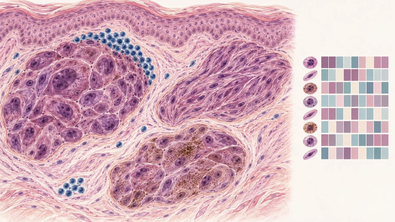
Separating cells also removes their original positions in the tissue. Spatially resolved methods retain location information, helping researchers see which cells sit near one another and how tumor and immune cells are arranged. A cell’s molecular state and its neighbors provide related but different clues: one concerns what the cell is doing, while the other concerns its place in the tissue’s interactions.
Combining single-cell and spatial analyses can support more detailed tissue atlases, suggest possible communication between nearby cells, and help trace how cell populations change. These are research opportunities described by the review. The methods have not been shown here to provide a validated way to select melanoma checkpoint treatment.
A biopsy captures one place at one time
A tumor is not necessarily uniform. Different cell populations can occupy different areas of one tumor, vary between disease sites, or change as the disease develops. The surrounding stroma and immune cells can vary too. A biopsy therefore gives a partial view from a particular place and moment, rather than a complete portrait of every tumor site and cell population in a patient.
Time adds another dimension. The review distinguishes differences present within a tumor from changes that emerge between an initial tumor and a later recurrence. Treatment can be followed by the growth of cell populations with different molecular features, contributing to resistance. Repeated biopsies during disease and treatment could show more of this evolution, but obtaining multiple samples is a concrete challenge. The review also discusses circulating tumor DNA as a possible way to monitor disease changes, while pointing to the need for more research.
Resistance can take different routes
The review separates primary resistance, when treatment does not produce an initial response, from acquired resistance, when a patient relapses after an earlier response. Tumors can become resistant through different changes, so a pattern found in one group need not describe another.
As a bounded example, the review cites a study of people with metastatic melanoma whose disease acquired resistance to anti-PD-1-based treatment. In that cohort, 28% had tumor PTEN loss and 22% had impaired HLA-A regulation. The two reported changes point to different possible routes to resistance across patients. The figures describe that cited cohort, not universal rates or a way to predict an individual’s course.
Treatment choices also involve trade-offs
A more detailed account of tumor and immune-cell variation could eventually help researchers understand why responses differ. Any useful clinical prediction would also need to help with actual choices, including the balance between treatment benefit and harm.
The review describes CheckMate 067, in which combined nivolumab and ipilimumab showed greater efficacy than either drug alone, alongside higher rates of immune-related adverse events. The comparison illustrates why treatment decisions are not only about finding a regimen with stronger activity. A dependable prediction could be valuable if it helped identify who might benefit from a more intensive option and who might avoid its added risks. The review’s discussion of cell mapping does not establish that it can yet make that distinction.
Where this research could lead
Imagine a person whose first melanoma biopsy shows little PD-L1. If future evidence supports it, clinicians could discuss treatment options using a dependable picture of immune-cell states and tumor neighborhoods across samples, rather than leaning on that reading alone. Existing checkpoint treatments would remain the starting point; the new capability would be a validated map that adds useful prediction. The low-PD-L1 response exception helps explain why such a broader picture may matter, while single-cell and spatial analyses offer complementary ways to build it.
A nearer milestone is prospective evidence that these maps predict response or relapse beyond existing markers across patients. Reliable sampling, consistent analysis, and clinical validation would all be needed before a map could support treatment selection. The review presents a direction for research, not evidence that this future capability is already available.
Key Sources
- Primary source: Unravelling Tumour Microenvironment in Melanoma at Single-Cell Level and Challenges to Checkpoint Immunotherapy.
DOI or Journal Page · Review or Meta-Analysis · 2022-09-28
Keep exploring