
Research Signal – Source date 2026-02-04
The same cell state can serve different purposes
Metabolic dysfunction-associated steatohepatitis (MASH), also called inflammatory fatty liver disease, involves fat accumulation, liver-cell injury, inflammation and scarring. In some people, progressive scarring can lead to cirrhosis and liver cancer. Hepatic stellate cells are central to this process: when activated, they produce components of the extracellular matrix, the material that builds up as scar tissue.
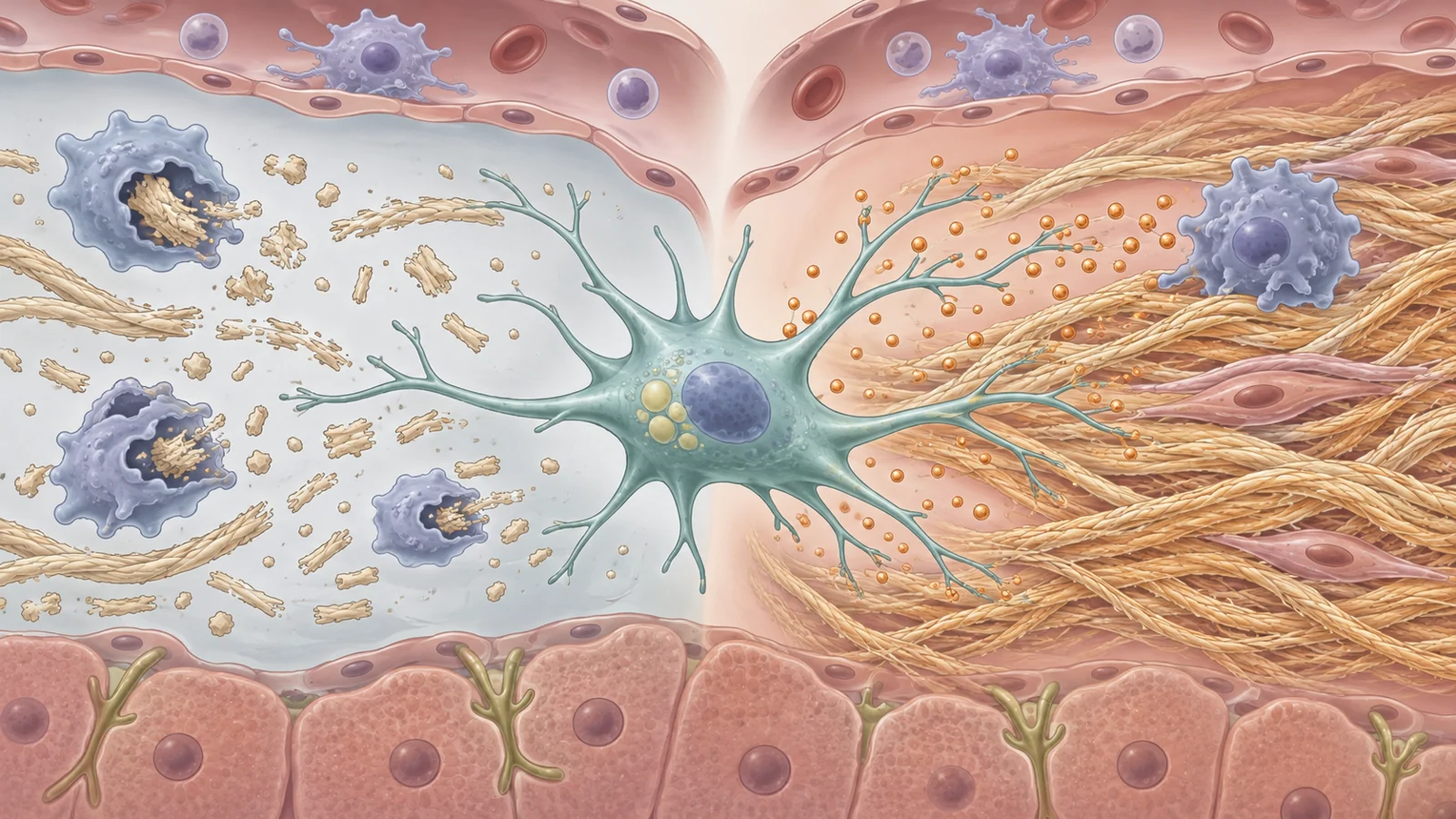
A review published on February 4, 2026, examines how senescence in these cells may shape MASH. Cellular senescence is a state in which a cell stops dividing. In stellate cells, that pause can curb the growth of scar-forming cells. But senescence also changes what a cell releases into its surroundings. The effects depend on the cell’s behavior and its environment, so the label “senescent” alone does not tell whether a stellate cell is helping tissue recover or sustaining disease.
In a short-lived response to injury, senescent stellate cells may limit scarring in several ways. They stop proliferating, reduce production of matrix components and release enzymes that break down deposited matrix. They can also attract immune cells that clear the senescent cells and help remodel scar tissue. In an acute repair setting, some of their secretions may support the growth of neighboring liver cells as the organ restores tissue.
The problem described in the review arises when metabolic and inflammatory stress persists. Senescent stellate cells may remain in the liver and develop an expanded senescence-associated secretory phenotype, or SASP. This means they release a changed mix of signaling molecules. Instead of helping resolve a temporary injury, that ongoing output can recruit inflammatory cells, activate other stellate cells and add to tissue stress.
Fat stress can reach stellate cells through vesicles
The review describes several pressures that may push stellate cells toward senescence, including lipotoxicity, oxidative stress, DNA damage, mitochondrial dysfunction and impaired autophagy. Autophagy is a cell’s process for removing or recycling damaged components; when mitochondrial upkeep falters, cell stress can compound. These routes are part of a proposed chain linking metabolic strain to altered stellate-cell behavior.
One example starts with fat-stressed hepatocytes, the main cells of the liver. In a high-fat-diet mouse model, small extracellular vesicles released by fatty, stressed hepatocytes carried a protein called LIMA1. After uptake by stellate cells, the vesicles were associated with increased LIMA1 in those cells, reduced PINK1-related mitochondrial autophagy and greater stellate-cell activation. This experiment offers a concrete possible route by which stress in one liver cell type can affect another. It is preclinical evidence from mice, not proof that this exact sequence drives disease in people with MASH.
Oxidative stress can contribute to the same broader picture. Reactive oxygen and nitrogen species can damage cellular components, including DNA. A sustained DNA-damage response is one mechanism associated with cellular senescence, while dysfunctional mitochondria can produce further oxidative stress. The review presents these interacting pressures as possible contributors to a state that may persist under chronic metabolic injury.
Secretions can relay stress across the liver
A senescent stellate cell’s secretions can influence more than its immediate surroundings. The review describes SASP factors such as CCL2, also called MCP-1, and CXCL1 as signals that can draw circulating monocytes into the liver. There, these cells may become macrophages with inflammatory or pro-fibrotic behavior. The review also discusses IL-1β and IL-6 as factors involved in that environment.
Macrophages can then send signals back. Activated macrophages release factors including TGF-β, PDGF and TNF-α, which can activate otherwise quieter stellate cells and support the survival of activated or senescent ones. This exchange could help maintain a cycle in which immune activity and scar-forming activity reinforce one another. The review discusses MASH-associated macrophage subsets in this context, while describing the influence of senescent stellate cells on those populations as an area of investigation.
The effects can extend to hepatocytes as well. In the pathway described by the review, IL-6 from the SASP can activate JAK/STAT3 signaling in nearby liver cells. Persistent signaling is linked in the reviewed research to mitochondrial dysfunction and DNA-damage markers in hepatocytes, potentially adding to injury and inflammation. These mechanisms help explain how a cell state in stellate cells could affect the wider liver environment; they do not establish that any one signaling route determines an individual patient’s course.
Immune clearance can help, but timing matters
The liver has ways to remove senescent stellate cells. The review describes natural killer (NK) cells recognizing ligands displayed by these cells, including molecules that interact with the NK-cell receptor NKG2D. Certain T cells can also promote stellate-cell death through Fas–FasL signaling. When immune surveillance clears senescent cells promptly, it may limit their continued presence and secretions.
But the target cell is not simply harmful in every setting. In an acute liver-regeneration experiment discussed by the review, senescent stellate cells released IL-6 and CXCR2-binding signals such as CXCL1 and CXCL2. These signals promoted neighboring hepatocyte proliferation through STAT3 and YAP-related pathways. Specifically eliminating the senescent stellate cells during that early regenerative phase impaired liver regrowth in the experimental setting.
That result makes indiscriminate cell removal a consequential choice. Clearing persistent cells might reduce damaging secretions, while removing cells that are supporting an early repair response could interfere with regeneration. The reviewed models and observations do not establish when, or how extensively, senescent stellate cells should be removed in people with MASH.
Treatment strategies face different tradeoffs
The review discusses two broad approaches. Senolytics aim to eliminate senescent cells. In preclinical work it summarizes, the combination of dasatinib and quercetin reduced senescent stellate cells, liver fat and TGF-β1 in experimental models, including a medaka fish model. Another senolytic, ABT-263, cleared senescent stellate cells and hepatocytes in a mouse liver-injury model and was associated with lower SASP-factor expression and improved mitochondrial function.
Those findings come with concerns. The review notes thrombocytopenia associated with senescent-cell removal and reports tumor-promoting effects of dasatinib and quercetin in hepatocellular-carcinoma models. These observations make clear that clearing cells across tissues or stages could have costs. They do not show that senolytics improve MASH outcomes in patients, or that the approaches discussed prevent liver cancer.
Senomorphics take a different approach: rather than remove senescent cells, they aim to change their secretions or behavior. The review discusses possible targets such as mTOR, p38/MAPK and regulators of NF-κB. It also notes that drugs including rapamycin, ruxolitinib, glucocorticoids and metformin have been considered for senescence-phenotype regulation. Because these pathways also serve normal cell functions, changing them could affect healthy cells as well. The potential tradeoff is therefore different, not automatically safer: dampening harmful secretions may leave the cells in place, but broad pathway effects remain a concern.
Finding the right cells remains a challenge
A targeted intervention would depend on identifying which cells are senescent, what they are secreting and whether they are supporting repair or sustaining chronic inflammation. No single marker described in the review resolves all of those questions. The cell-cycle regulator p16INK4a, for example, can be measured in tissue, but a marker of senescence does not on its own reveal the cell’s current effects on surrounding tissue.
Blood measurements pose another difficulty. Senescence-associated secretions can be detected in plasma, but the review notes that they may also reflect inflammation elsewhere in the body. It discusses senescence-related genes in circulating T cells and acyl-CoA-binding protein as possible indicators under study, not as a validated way to select people with MASH for treatment.
Imaging has limits too. Elastography can assess fibrosis, but the review describes challenges in early disease and in identifying senescent stellate cells specifically. PET tracers aimed at senescence-related activity are described as promising in preclinical work, with early human research mentioned. The examples do not establish a validated MASH-specific test for deciding who should receive a senescence-targeted therapy.
What selective treatment could change for MASH
If researchers can distinguish stellate cells that are sustaining chronic inflammatory signaling from those participating in repair, future treatments might reduce harmful scarring and inflammation while preserving the liver’s capacity to recover. Reaching that possibility would require reliable ways to identify the relevant cells and evidence that a carefully timed intervention improves outcomes safely in people with MASH. The February 2026 review organizes biological reasons to investigate that approach; it does not establish a patient treatment or a clinical selection method.
Key Sources
- Primary source: Senescent hepatic stellate cells drive inflammation and disease progression in MASH (Review).
DOI or Journal Page · Review or Meta-Analysis · 2026-02-04
Keep exploring