
Research Signal – Source date 2022-09-21
Age is the strongest established risk factor for age-related macular degeneration, but aging alone does not identify the cellular events that damage sight. One possibility is cellular senescence, a state in which cells undergo persistent stress-related changes and can release signals that affect their surroundings.
That possibility creates an appealing treatment idea. If senescent cells help sustain harmful inflammation, drugs that remove them might protect the aging eye. But retinal tissues contain cells that divide very little or not at all. Removing a damaged cell may also remove a piece of a support system that is difficult to restore.
A review published on September 21, 2022, assessed whether senescence might contribute to the development or progression of age-related macular degeneration. Its conclusion was cautious. The biology is plausible, and several observations justify further study, but the evidence does not establish senescence as a causal driver of human macular degeneration.
Why the retinal pigment epithelium changes the question
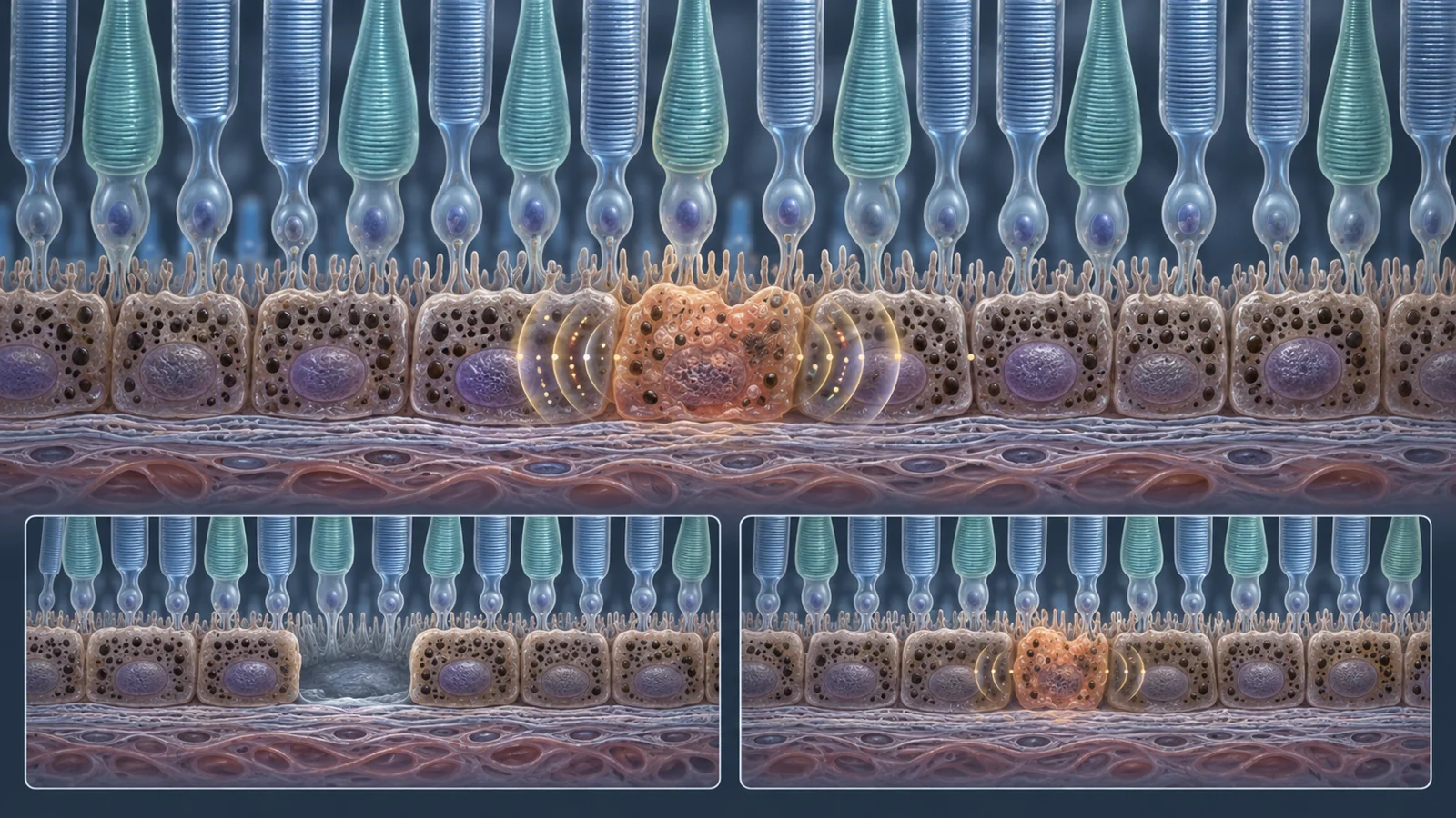
The retinal pigment epithelium is a thin support layer beneath the light-sensitive photoreceptors. It helps move nutrients and ions, process metabolic waste, regulate the environment around photoreceptors, and maintain the blood-retina barrier. It also handles photoreceptor outer segments and produces factors that help preserve photoreceptor integrity.
Those jobs depend on organization. An RPE cell is not simply a generic cell occupying space; its upper and lower surfaces have different structures and functions. The layer must maintain its contacts with neighboring cells and regulate what crosses between the retina and the underlying tissue.
Senescence was originally described as an irreversible loss of the ability to divide, often accompanied by resistance to cell death. More recent work has suggested that senescence-like states can also occur in cells that have already stopped dividing, including neurons. That matters in the eye because mature retinal neurons and RPE cells are largely post-mitotic. A change in a non-dividing cell cannot be interpreted in exactly the same way as a change in a cell that has reached the end of repeated replication.
The review describes senescence as a broader cellular state, not merely a failure to divide. Senescent cells can produce a changing mixture of inflammatory cytokines, chemotactic signals, and damage-associated molecules known collectively as the senescence-associated secretory phenotype. Those secretions may influence neighboring cells, alter tissue behavior, or sometimes participate in useful processes such as wound healing. The effect depends on the cell type, tissue, trigger, and surrounding environment.
A cell culture can ask the wrong biological question
A large part of the difficulty begins before a marker is measured. Many experiments on RPE senescence have used ARPE-19, an immortalized RPE cell line that can remain active in the cell cycle and is often poorly differentiated. In that setting, an experiment may be studying stress in dividing cells rather than the biology of mature RPE in the eye.
A more faithful RPE model is non-dividing, polarized, and visibly differentiated. Here, polarized means that the cell layer has an organized top side and bottom side, with proteins and transport functions distributed according to their position. That arrangement is tied to barrier performance and to how the layer responds to oxidative or complement-related stress.
Primary RPE cells from human or animal tissue can reproduce several features of the tissue when grown as organized monolayers on permeable supports. The review also describes protocols that improve the phenotype of ARPE-19 cultures. Even those improved approaches do not erase every limitation. The cells may still have a relatively weak barrier and chromosomal abnormalities, and their response to stress may differ from that of mature RPE in a human eye.
The experimental starting point therefore changes the question. A result in actively dividing ARPE-19 cells may show that a particular insult can produce a senescence-associated response under those culture conditions. It does not automatically show that the same insult creates the same state in mature human RPE or that the resulting cells contribute to macular degeneration.
A marker is a clue, not a diagnosis
Researchers often begin with markers such as SA-β-gal activity or increased expression of p16INK4a and p21CIP1. Other useful observations can include signs of DNA damage, changes involving regulators of inflammatory secretion, and shifts in bioactive lipids. The review emphasizes that several markers are more informative together than any one measurement alone.
SA-β-gal staining, for example, reflects increased lysosomal activity that often accompanies senescence-related stress. But aging cells can accumulate long-lived lysosomes for reasons that are not equivalent to a harmful senescent state. Similarly, increased p16 or p21 expression does not always indicate senescence; the review specifically notes that p16 can also mark inflammation and general aging. A positive signal does not by itself establish that a cell is senescent, dangerous, dispensable, or suitable for removal.
Observations in aging tissue illustrate the problem. Increased SA-β-gal staining has been seen in human and non-human-primate RPE with age, but the review presents the interpretation as unresolved. The staining could reflect senescence or increased reactivity associated with long-lived lysosomes in post-mitotic cells; the review leaves that distinction unresolved.
Animal models provide another kind of clue. Aged SAMP8 mice have been described with ocular autofluorescence, reduced retinal function, deposits beneath the RPE, degenerative changes, and increased p16 expression in RPE cells. In a separate mouse experiment, sub-retinal exposure to amyloid beta was associated with changes in retinal electrical responses, RPE degeneration, and increased p16 expression. These findings make senescence-related mechanisms worth investigating, but they do not demonstrate that senescent cells cause human age-related macular degeneration. The disease context, the cell type, and the meaning of each marker still have to be connected carefully.
For human AMD, the central issue is not whether a single signal can be detected. It is whether a defined population of cells shows a consistent, multi-marker senescence profile in the relevant tissue, whether that population changes with disease stage, and whether its behavior plausibly contributes to loss of retinal function.
How stressed cells could affect their neighbors
The proposed mechanism is partly about communication. A persistently stressed cell can release inflammatory cytokines, chemokines that attract or guide other cells, and damage-associated signals that alert nearby tissue. Through this secretory activity, a relatively small population could influence the behavior of surrounding cells without directly replacing them or becoming the only source of damage.
In the eye, such signaling could matter because the RPE, photoreceptors, choroid, and immune-related cells operate as a connected system. The RPE helps maintain the conditions photoreceptors need, while photoreceptors place continual metabolic demands on their support layer. A change in one compartment may therefore alter the environment experienced by the others.
The same biology also argues against treating every stressed cell as an enemy. Senescence-associated secretions are dynamic, and some forms of senescence can have beneficial roles in repair. A cell that expresses one associated marker may still be contributing to tissue structure or performing a function that neighboring cells cannot readily replace. The important therapeutic target would be a harmful, persistent state in a relevant cell population, not simply any old or stressed cell.
Removing cells is not the only therapeutic option
Senolytics are drugs intended to eliminate senescent cells. Their appeal is direct: if a small population is maintaining damaging signals, clearing it could reduce the source. But retinal tissue raises a specific cost. Photoreceptors and other retinal cells have limited capacity for replacement, and RPE cells provide essential support even when they are no longer dividing. Removing cells could weaken the tissue if the population is larger, more functional, or more structurally important than expected.
Senomorphics, sometimes called senostatics, take a different approach. Instead of removing the cell, they aim to reduce selected harmful parts of its secretory program. This could preserve a living support cell while dampening signals that affect its neighbors. The tradeoff is that secretion suppression may require continued treatment, and the undesirable signals could return when treatment stops. The approach also depends on being able to separate harmful signaling from functions the cell still needs to perform.
The review notes that the Bcl-xL inhibitor UBX1325 was being tested in a phase 2a human trial for refractory diabetic macular edema and neovascular age-related macular degeneration. That mention provides contemporary context for the therapeutic direction; it is not evidence of improved vision or established efficacy for AMD. The review also highlights unresolved questions about treatment duration, dosing intervals, tissue atrophy, and the risk of affecting cells that are difficult to replace. A drug already used or investigated for another condition would still require evidence specific to this ocular use.
The practical decision is therefore more demanding than finding p16-positive cells. Researchers would need to show that the targeted cells contribute to disease, that the intervention reaches the relevant tissue, and that removing or quieting them preserves the RPE’s barrier and support functions. Those requirements connect cell identification to tissue integrity rather than treating them as separate problems.
What targeting senescence could mean for preserving vision
A future treatment could become useful for people with macular degeneration if it reduces damaging senescence-associated signals while retaining enough retinal support to protect remaining photoreceptors. In another version, carefully removing a defined harmful cell population might slow tissue deterioration without creating a new structural deficit.
That benefit remains conditional. It depends on evidence that senescence contributes to the relevant form of human macular degeneration and that the chosen intervention can distinguish harmful targets from cells that are old, stressed, or still doing essential work. The goal would be preservation of remaining visual function, not replacement of lost retinal cells or reversal of established vision loss.
The review’s central question is therefore still open, but it is becoming more precise. Progress will depend on matching the right cell models to mature RPE biology, identifying senescence with multiple lines of evidence, and testing whether changing that state protects the tissue that sight still depends on.
Key Sources
- Primary source: Does senescence play a role in age-related macular degeneration?
DOI or Journal Page · Review or Meta-Analysis · 2022-09-21
Keep exploring