
Research Signal – Source date 2024-06-01
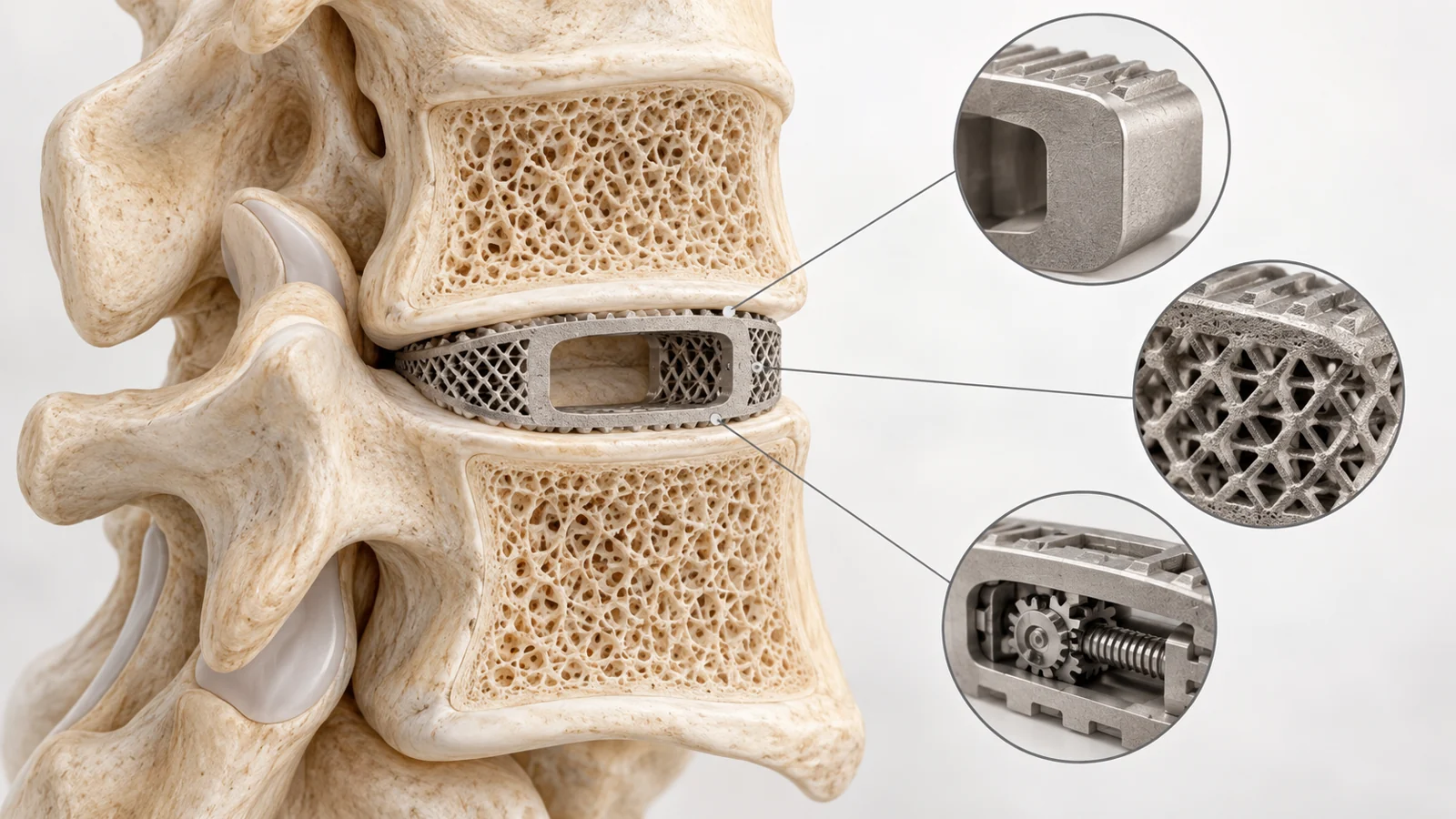
A spinal-fusion cage has to do several jobs in a small space. Placed between vertebrae in the lower back, it provides structural support and room for bone to grow across the space as the bones fuse. It must withstand loading, fit through the surgical access route, and interact with the bone supporting it. Its material also affects how clearly clinicians can assess fusion on postoperative images.
Those demands can pull in different directions. A strong metal may be much stiffer than surrounding bone. A plastic that makes imaging easier may have a surface that does little to encourage bone attachment. A cage that fits well once positioned may still be difficult to insert.
A narrative review published in June 2024 brings these competing demands together, examining materials, porous structures, coatings, biodegradable composites, and expandable cages. It describes increasingly sophisticated ways to solve specific engineering problems. Whether those changes consistently improve pain, disability, or fusion outcomes remains a separate question, especially in comparisons of expandable and fixed-size devices.
Every material changes the compromise
Titanium and its alloys offer strength, corrosion resistance, and compatibility with living tissue. Their ability to support bone ingrowth makes them useful candidates for a device intended to become integrated with the spine. But strength and stiffness are different properties. Stiffness describes how much a material deforms under a load, and conventional titanium cages can be substantially stiffer than native bone.
That mismatch affects how forces are shared. If an implant carries too much of the load, adjacent bone can experience less loading, a problem called stress shielding. The review also discusses subsidence, in which a cage sinks into the supporting vertebral bone and may compromise the restored space between vertebrae. These are distinct concerns about the relationship between the implant and its surroundings. Titanium also obscures parts of postoperative images, making the developing fusion harder to assess.
Polyetheretherketone, usually shortened to PEEK, is an implant plastic with a different balance of properties. Its stiffness is closer to that of cortical bone, the dense outer portion of bone, and its relative transparency on imaging helps clinicians see around the implant. Those features address load transfer and visibility. Its biological interface is less favorable, however. PEEK is relatively resistant to wetting and biologically inert, giving researchers a reason to modify its surface to encourage tissue attachment.
Tantalum adds another option. The review highlights its corrosion resistance and compatibility with tissue, particularly in porous forms whose architecture and stiffness resemble cancellous bone, the spongy bone inside vertebrae. It also discusses tantalum applications elsewhere in orthopedics and in cervical spine surgery. Those examples help explain interest in the material without making it a universal replacement for titanium or PEEK in lumbar fusion.
Material names therefore describe only part of a cage’s behavior. The solid substance matters, but so do the spaces built into it and the surface that bone encounters.
Building the interface into the cage
Three-dimensional printing allows manufacturers to create intricate porous structures within a cage. This changes more than its appearance. The arrangement of solid material and open space can alter the device’s effective stiffness while providing routes for bone ingrowth and, potentially, blood-vessel development.
The review describes printed titanium structures with stiffness closer to native bone than conventional titanium-alloy cages. This is an architectural solution to a material problem. Titanium remains titanium, but organizing it into a porous structure changes how the implant responds to force. The design challenge is to preserve adequate support while creating a structure suited to biological integration.
Surface modification works at the boundary where implant and tissue meet. A cage can retain a load-bearing core while its outer surface is made porous or coated with another substance. For PEEK, the reviewed approaches include titanium, hydroxyapatite, and calcium silicate modifications intended to increase biological activity. Hydroxyapatite is a mineral used in these composites and coatings to help create a more favorable tissue interface.
Laboratory findings help show what such changes are trying to accomplish. The review describes work in which porous PEEK supported greater cell proliferation and cell-mediated mineral deposition than smooth PEEK and titanium. These observations concern how cells respond to a surface. They provide a mechanism to investigate, rather than a direct measurement of recovery after lumbar surgery.
The interface must also survive implantation. In testing summarized by the review, porous PEEK devices sustained little damage during aggressive cervical insertion impacts, while titanium-coated PEEK devices lost a substantial portion of their initial coating coverage. Although that testing concerned cervical devices, it illustrates a concrete engineering constraint: a carefully designed surface needs to remain attached when the cage is inserted. Coating composition, pore structure, and resistance to impact are connected parts of the same design problem.
A disappearing cage must keep its strength long enough
Biodegradable cages introduce a timing requirement. A permanent implant can continue providing support after fusion. A resorbable implant is intended to gradually disappear and ideally be replaced by natural bone. For that approach to work, the loss of implant strength must stay compatible with the development of a stable fusion.
Polylactic acid, or PLA, illustrates why that timing matters. The review discusses a systematic review of biodegradable implants, predominantly polylactides, that found insufficient support for routine use of absorbable cages because of unfavorable long-term fusion results. Early degradation and loss of strength were proposed explanations for poorer outcomes, including bone loss and accelerated subsidence. The problem is therefore more specific than whether a material can dissolve safely. It must retain enough mechanical function throughout healing.
Polycaprolactone, or PCL, is another biodegradable polymer explored in composites with calcium-based ceramics. Combining materials offers ways to adjust stiffness, degradation, and biological activity together. The review describes spinal-fusion experiments in pigs and sheep using PCL-based scaffolds, including combinations with calcium phosphate and bone-growth-promoting substances.
In one sheep cervical-fusion study summarized by the authors, a PCL–tricalcium phosphate scaffold supported bone ingrowth and maintained load-bearing capacity, with fusion efficacy comparable to titanium cages containing the animal’s own bone graft. That is an animal result in the neck, not a demonstration of equivalent outcomes in human lumbar surgery. Its specific contribution is to show how a degradable composite might combine temporary support with tissue growth.
The central challenge remains coordinating two processes that change over time. Bone must take over the structural role as the scaffold loses it. Adjusting mineral content can make a polymer composite stiffer, but the complete device still has to maintain support through the healing period.
Expanding inside the spine solves an access problem
Static cages have a fixed shape and size. Expandable cages can enter the surgical space with a smaller profile and enlarge after placement. That creates a practical attraction: the dimensions needed for insertion do not have to be identical to the dimensions needed for support.
The review describes expansion as a way to adjust fit and reduce the forceful insertion sometimes required for a static cage. Avoiding those impacts could help protect the vertebral endplates, the bone surfaces supporting the device. Expansion can also change the height of the space between vertebrae and the alignment at the treated spinal segment.
The surgical route matters. Approaches from the front or side can accommodate cages with a larger contact area than some approaches through the back. The review reports favorable subsidence findings for expandable cages in some lateral procedures, where avoiding forceful insertion may be especially relevant. It also describes conflicting findings in transforaminal lumbar interbody fusion, an approach through the back and toward one side of the spine. Two meta-analyses of that procedure found no significant subsidence difference between expandable and static cages.
Imaging results are similarly uneven. Some comparisons favored expandable cages for restoring disk-space height or local curvature. A meta-analysis, however, found no statistically significant difference in several measures of spinal alignment. A device can make a particular adjustment easier without producing a consistent advantage across all anatomical measurements.
The patient-centered comparisons provide the clearest limit on claims of superiority. Three meta-analyses summarized in the review found no significant differences between expandable and static cages in back and leg pain scores, disability scores, or fusion rates for the transforaminal procedure. These findings do not prove that every design performs identically in every setting. They do mean that improved fit or a favorable postoperative image cannot, by itself, establish better recovery. The narrative review brings together these comparisons; it does not validate a new cage through its own clinical trial.
What this could change
The future capability described by this work is a cage whose shape, mechanical behavior, and biological interface can be tailored together. Patient-specific geometry could accommodate an individual’s anatomy. A porous structure could adjust how loads pass between implant and bone. A durable surface could encourage bone attachment, while locally delivered growth factors or antibiotics could add functions beyond structural support.
If those features remain effective through insertion and healing, and translate into clinical benefit, spinal-fusion implants could offer a more individualized combination of fit, support, and local treatment. A successful resorbable version could eventually leave natural bone carrying the load without a permanent cage. The practical payoff would come from coordinating those functions over the course of recovery, so that the device’s changing role matches the tissue’s changing needs.
Key Sources
- Primary source: Innovative Developments in Lumbar Interbody Cage Materials and Design: A Comprehensive Narrative Review.
DOI or Journal Page · Review or Meta-Analysis · 2024-06-01
Keep exploring