
Research Signal – Source date 2026-06-01
A transplant can give a child years of life that severe illness might otherwise take away. Protecting that future involves a difficult balance between controlling harmful immune reactions and preserving defenses against infection and cancer. A review published June 1, 2026, examines another dimension of that balance: how transplantation and its associated treatments can change the immune system’s capacity to renew itself.
The authors bring together evidence that some pediatric transplant recipients develop immune features usually associated with older adults. These include fewer inexperienced T cells, a narrower range of immune recognition and persistent inflammatory activity. The term immunosenescence describes aging-related changes in immune defenses. Here, it concerns particular cells and functions, without implying that a child’s whole body has become elderly.
The review’s contribution is to connect these mechanisms with patterns of cancer after childhood transplantation and the practical requirements for testing that connection. Its central question is consequential: could preserving the quality of immune recovery help protect children throughout years of transplant survivorship?
Rebuilding cell numbers can leave gaps in immune coverage
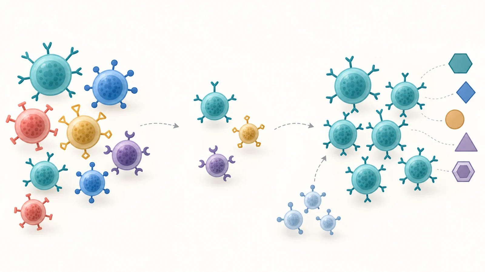
T cells help coordinate immune responses and recognize infected or abnormal cells. Their diversity matters because different T cells recognize different targets. Naive T cells have not yet encountered their matching target; memory populations have already been shaped by previous encounters. A recovering immune system needs more than an adequate total cell count. The range of cells that returns also matters.
The review describes how anti-thymocyte globulin, or ATG, can alter that rebuilding process. Used in some transplant regimens, this antibody treatment depletes T cells. The findings summarized by the authors indicate that naive populations can be especially affected while some memory populations are relatively spared. Recovery may then depend heavily on surviving cells dividing to replenish the depleted population.
That route can restore numbers while producing many descendants of a more limited set of cells. Meanwhile, the thymus—the organ where new T cells develop—may have been injured by conditioning treatment or surgery. If new-cell production is constrained, peripheral expansion cannot necessarily restore the breadth of recognition that was lost. Repeated division can also shorten telomeres, the protective structures at chromosome ends.
Persistent stimulation adds another pressure. The authors identify cytomegalovirus, or CMV, reactivation as one influence that can drive immune populations toward highly differentiated states. Ongoing responses to the transplant and sustained inflammatory signaling can contribute as well. The resulting profile may include loss of the T-cell marker CD28, increased expression of markers such as KLRG1, and changes in function.
These observations describe related but distinct processes. Cellular senescence involves a durable arrest of cell division; exhaustion concerns impaired function under persistent stimulation; a shift from naive to memory cells describes population composition. They can coexist, but one marker cannot make them interchangeable. The proposed cancer connection also has two parts: reduced capacity to recognize abnormal cells, and an inflammatory environment that may help those cells grow. Senescent cells can release inflammatory molecules, growth factors and other signals, potentially linking impaired immune renewal with conditions favorable to tumors.
Reading the cancer figures without turning them into a prediction
The epidemiology summarized in the review shows substantial excess cancer after pediatric transplantation. Understanding the figures requires keeping the population, diagnosis and observation period together.
One US registry analysis covered 17,958 solid-organ transplants performed before age 18, with a median follow-up of four years and a maximum of 22. The review reports an all-cancer standardized incidence ratio of 19.1. This compares observed cancers with the number expected from population rates. It does not mean that 19.1% of recipients developed cancer, or supply a lifetime probability for a particular child. A large relative increase can arise when the expected number in the comparison population is small.
The same table reports a ratio of 212 for non-Hodgkin lymphoma and post-transplant lymphoproliferative disease, or PTLD, a group of conditions involving abnormal lymphocyte growth. Risks varied greatly by transplanted organ, with particularly high relative figures in intestinal and heart or lung recipients. The early peak after transplantation is consistent with intense immunosuppression and Epstein–Barr virus, or EBV, infection in children without prior exposure. This EBV-associated lymphoproliferative risk is distinct from CMV’s role in reshaping immune populations.
A separate stem-cell transplant cohort illustrates why the headline numbers cannot be combined into one general estimate. As summarized by the review, it included 6,028 recipients younger than 21 who underwent allogeneic transplantation for nonmalignant diseases. There were 71 subsequent neoplasms, reported as 1.2%, with a median follow-up of 7.8 years; the standardized incidence ratio was 11. PTLD was excluded from that analysis’s definition of subsequent neoplasms. The observed percentage and the relative ratio answer different questions, and neither supports a direct ranking against the solid-organ cohort, whose age limits, follow-up and cancer endpoints differed.
Timing changes the picture further. Early lymphoproliferative disease accounts for an important part of the pediatric cancer burden, while solid tumors become more prominent with longer follow-up into adolescence and adulthood. In stem-cell transplant survivors, prior treatment and graft-versus-host disease can contribute to malignancies emerging years later. A single multiplier compresses these different histories too severely to describe an individual child’s situation.
Separating immune changes from other causes
Excess cancer establishes a problem to explain. It does not determine how much of that problem is caused specifically by immune aging.
Some children, particularly those receiving stem-cell transplants for cancer, have already received chemotherapy or radiation. Such exposures can damage DNA and independently increase later malignancy risk. Some inherited disorders that lead to transplantation also carry cancer susceptibility through genomic instability or defective DNA repair. These influences can coexist with immune depletion, viral exposure and impaired immune renewal; they cannot simply be assigned to a single transplant-related mechanism.
Detection differs too. Transplant recipients undergo intensive medical follow-up, creating more opportunities to discover otherwise unnoticed tumors. The authors discuss this surveillance effect as a possible contributor to observed rates of cancers such as thyroid and renal tumors. It does not erase the excess burden, but it affects its interpretation.
To establish the independent contribution of immune aging, studies would need to connect immune trajectories with later outcomes while accounting for these competing explanations. The mechanisms assembled in the review provide reasons to investigate that connection. The cohort summaries do not quantify what fraction of cancers those mechanisms cause.
When an immune measurement becomes useful
The authors discuss several ways to examine immune aging. Flow cytometry can identify cell populations through combinations of surface markers. Telomere measurements can probe one aspect of cellular replication history. Epigenetic clocks use patterns of DNA methylation to estimate aspects of biological aging. Each captures a different dimension, so a result requires interpretation before it can inform care.
Children make that interpretation especially demanding. Expected immune profiles change across infancy, childhood and adolescence, and infection and vaccination history also matter. A marker frequency that looks unusual against an adult reference might have a different meaning in a growing child. The review therefore emphasizes age-specific reference ranges, with CMV history and treatment exposures recorded alongside the measurements.
Repeated observations can be more informative than a single sample. A result soon after T-cell depletion, viral reactivation or treatment for rejection may reflect a temporary disturbance. The authors propose tracking changes against a patient’s baseline and major clinical events, looking for persistent departures from age-adjusted expectations. That approach could distinguish a sustained loss of naive cells from an isolated fluctuation. It remains a proposed interpretation strategy, rather than an established cancer-prediction threshold.
Reproducibility is another practical requirement. Laboratories need harmonized assays and reporting so that a change in measured cell populations reflects biology rather than differences in testing. The authors identify flow cytometry as a relatively implementable starting point in specialist pediatric transplant centers, while telomere and methylation assays may require additional laboratory resources. Even a reproducible test must then show that it predicts meaningful outcomes and adds useful information beyond a child’s existing clinical history.
Changing the trajectory brings a transplant safety tradeoff
Measuring a potentially harmful process and safely changing it are separate achievements. The review explores interventions that would act at different points in immune recovery, including clearing senescent cells, supporting the production of new T cells and altering metabolic pathways. These are directions for clinical evaluation, not demonstrated methods of preventing cancer in pediatric recipients.
Senolytic drugs aim to remove senescent cells. In principle, reducing a source of inflammatory signals might improve the environment in which immune cells function. But clearing cells would not by itself guarantee restoration of a diverse T-cell population. Any transplant application would have to establish which cells are affected and whether the net result preserves useful defenses.
Supporting immune renewal addresses another part of the problem. The authors discuss thymic regeneration strategies and proposed studies of interleukin-7, a signaling molecule involved in T-cell biology. Their suggested outcomes include measures of new-cell production, diversity of T-cell recognition and rejection. That combination captures the governing constraint: stronger immune activity must remain compatible with transplant safety. For solid-organ recipients, restored immune responsiveness could also include responses against the graft.
The proposed studies of senolytics, immune renewal and metabolic interventions should therefore be read as a research agenda. Improvements in a laboratory marker would be an intermediate result. Demonstrating benefit would require connecting those changes to better health while accounting for rejection, harmful immune reactions, infection and other treatment risks.
What this could change
If repeated immune measurements can identify trajectories that predict later illness, transplant teams could gain a more informative view of recovery than cell counts alone provide. If interventions can then safely alter those trajectories, that information could help tailor treatment and follow-up to preserve a broader, more durable set of immune defenses.
The potential benefit would extend across years of survivorship. Children could retain the function of a lifesaving transplant while facing fewer infections and a lower burden of later cancer, provided that preserving immune competence proves to improve those outcomes. The clinical advance would be the ability to protect long-term health without sacrificing the transplant that made that future possible.
Key Sources
- Primary source: Immunosenescence and cancer predisposition in pediatric transplant recipients: An emerging paradigm.
DOI or Journal Page · Peer-Reviewed Paper · 2026-06-01
Keep exploring