
Research Signal – Source date 2025-06-20
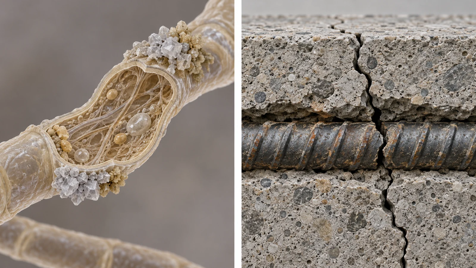
Concrete cracks can let water and corrosion agents reach the steel reinforcement inside a structure. A mineral that forms within a crack could, in principle, help close that route and extend the concrete’s useful life. Researchers are exploring whether fungi can encourage calcium carbonate (CaCO₃) to form, but a key question is what minerals those fungi actually produce—and how consistently they form across tiny regions.
A study published on June 20, 2025, examined deposits from three calcium-producing fungi with nanoscale X-ray methods. Calcite, a stable form of calcium carbonate, dominated the analyzed material. Yet the maps also revealed smaller contributions from other calcium forms, with clear variation among locations in one sample. The work characterizes fungal-grown minerals in culture; it did not test whether they seal a concrete crack.
The deposits formed in culture, not concrete
The researchers isolated three urease-positive fungi from environmental samples and grew them in a calcium-containing culture medium. Urease is an enzyme involved in breaking down urea; in this experiment, the fungi were selected for that activity and their ability to tolerate alkaline conditions. After incubation, the researchers collected the resulting mixture of fungal material and minerals.
To examine that mixture with X-rays, they prepared very thin sections: about 75 to 200 nanometers thick, depending on the fungal sample. An electron microscope showed the sections’ structure, while the X-ray measurements supplied chemical information. The researchers scanned multiple small regions rather than treating each deposit as a single, uniform mineral sample.
This distinction in scale matters. A bulk measurement can identify minerals present in a whole sample while averaging over different local conditions. Looking across tiny regions offers a way to see whether a mineral phase is spread throughout or concentrated in particular places.
Reading calcium and carbon signatures
The team used scanning transmission X-ray microscopy, or STXM, to image the sections at selected energies. At each energy, the amount of X-ray absorption varied with the elements and chemical forms in the material. Near the calcium and carbon absorption edges, those patterns also helped identify which forms were present. The resulting image stacks combined spatial information with spectra—chemical fingerprints collected at each point.
Calcium-edge spectra helped distinguish calcite from aragonite, another form of calcium carbonate. The same analysis also detected calcium signals that did not match carbonate. Those signals could not be reliably separated into calcium chloride versus calcium associated with extracellular polymeric substances (EPS), the materials fungi release around themselves. The study therefore grouped them as non-carbonate calcium rather than assigning a more specific identity.
Carbon-edge spectra offered a complementary check. Regions with calcium signatures consistent with calcite also showed carbon features consistent with carbonate minerals. In some locations, additional carbon features suggested contributions from organic material such as fungal polymers. This cross-check supports the interpretation of the analyzed deposits; it is not a test of how strongly a mineral would bind or how well it would repair concrete.
Calcite dominated, but one sample varied across five spots
Across analyzed sections from all three fungi, calcite was the predominant identified calcium carbonate phase. The researchers also found smaller amounts of aragonite and non-carbonate calcium signals. The overall pattern was consistent, but the composition was not identical at every location.
The clearest local variation appeared in five analyzed spots from fungal species 2. Three were dominated by calcite, with only minor contributions from other calcium forms. One showed mixed carbonate and non-carbonate calcium signals; another was predominantly non-carbonate calcium. These results show why an overall label such as “mostly calcite” can conceal meaningful differences within a deposit.
The maps locate those differences in the prepared samples. They do not show how a deposit changed over time, or establish that a particular mineral sequence occurred. Their contribution is a closer view of where the detected calcium forms were concentrated.
Local conditions may shape which minerals form
The authors suggest that conditions around fungal growth could help explain the observed patterns. Local differences in calcium availability, fungal activity, acidity, and the surrounding polymer-rich material may influence where carbonate minerals nucleate and which forms persist. EPS could also provide sites where calcium accumulates before carbonate forms.
One possible interpretation is that some calcium-rich regions first support less stable or intermediate mineral forms that later give way to calcite. The spatial snapshots are compatible with such a pathway, but they do not directly track a transformation. The proposed roles of metabolism and fungal polymers remain explanations to investigate, rather than causes demonstrated by these maps.
What the laboratory results leave open
The deposits were grown in culture and examined after preparation as ultrathin sections. The experiment did not investigate mineral formation in concrete or mortar, and it did not demonstrate crack sealing. It also did not measure recovered structural strength, corrosion prevention, service life, or emissions from concrete infrastructure.
The measurements have limits at this scale. Very low-density regions produced noisy spectra, and section thickness affected which regions could be analyzed. Fixation, washing, and other preparation steps may have altered fragile material around fungal cells, potentially affecting the carbon signatures and the apparent contribution of EPS. In addition, the calcium spectra could not distinguish calcium chloride from calcium associated with EPS in the grouped non-carbonate signal.
The authors identify testing in real concrete as necessary to assess how fungal mineral formation performs under the varied conditions structures face. That step would connect nanoscale composition to the practical questions the culture study leaves unanswered: whether minerals form where cracks occur and whether they remain useful within a cement-based material.
Why the mineral map could matter for concrete repair
If fungi can produce useful mineral deposits inside concrete cracks under real structural conditions, the approach could eventually help protect reinforcing steel and extend the service life of infrastructure. The predominance of calcite in these culture-grown deposits gives researchers a specific mineral outcome to investigate. But reaching that benefit depends on showing that the fungi and their deposits work in concrete, where local mineral formation must translate into a durable seal. This study provides a way to examine that formation at small scales; it does not yet show a repaired structure.
Key Sources
- Primary source: Nanoscale Characterization of Fungal-Induced CaCO 3 Precipitation: Implications for Self-Healing Concrete.
DOI or Journal Page · Peer-Reviewed Paper · 2025-06-20
Keep exploring