
Research Signal – Source date 2024-05-17
Some men who carry the fragile X premutation later develop fragile X-associated tremor/ataxia syndrome, or FXTAS, a progressive disorder involving movement and thinking. A 2024 MRI study asks whether a small anatomical feature around brain blood vessels might help explain why the disorder is associated with a particular pattern of changes.
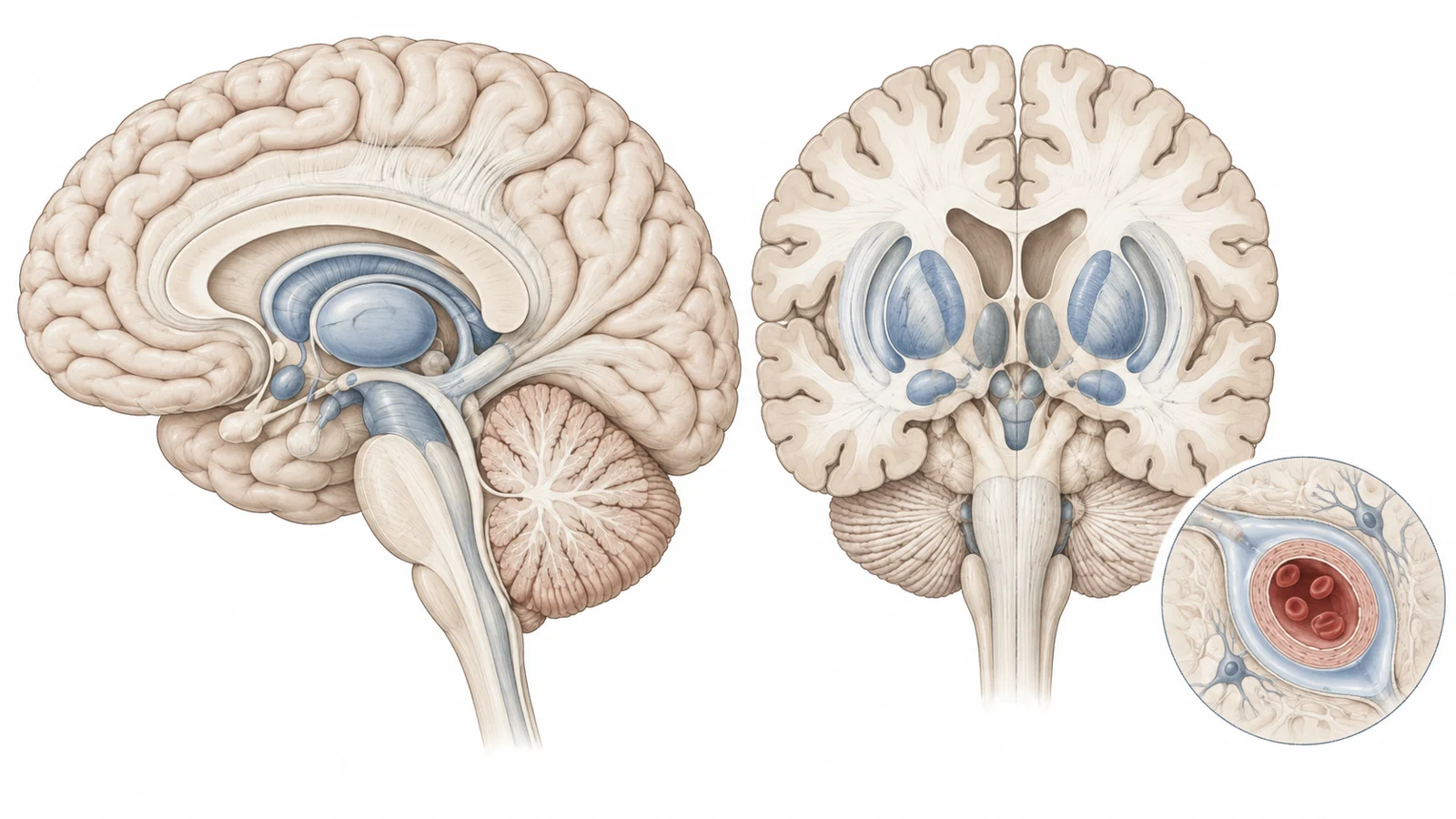
The feature is called an enlarged perivascular space. Perivascular spaces are fluid-filled passageways that surround small vessels in the brain. They can become visible on MRI, especially in areas such as the basal ganglia, deep white matter and midbrain. The researchers wanted to know whether these visible spaces were more prominent in male premutation carriers, whether their location mattered, and whether their ratings tracked clinical or other MRI measures.
The findings point to a regional association rather than a universal MRI signature. Spaces rated in the basal ganglia were more prominent in men with FXTAS and were related to several measures of motor, cognitive and structural change. But the study remains observational. It does not show that enlarged spaces cause impairment, directly demonstrate abnormal fluid circulation, or tell an individual carrier what will happen next.
Why these spaces are being studied
The fragile X premutation involves 55 to 200 CGG repeats in the FMR1 gene. Some carriers develop FXTAS later in life. MRI findings associated with the syndrome can include white-matter abnormalities, enlargement of the ventricles and generalized loss of brain volume. The middle cerebellar peduncle sign, a pattern of white-matter hyperintensity in pathways connecting the cerebellum with the brainstem, is one of its better-known radiological features.
Perivascular spaces are not the same as those larger-scale MRI findings. They are small fluid-filled spaces that follow the course of perforating vessels. On the scans used in this study, they appeared as sharply defined structures with the signal characteristics of cerebrospinal fluid. A space looked round when viewed across its short axis and linear when the scan followed its length.
The researchers rated spaces in three regions. The basal ganglia are deep brain structures involved in movement and other functions. The centrum semiovale is a region of deep white matter, while the midbrain is part of the brainstem. Ratings were based mainly on visible counts in the basal ganglia and centrum semiovale, with a simpler presence-based rating for the midbrain. The worse hemisphere was used when the two sides differed.
This matters because the same MRI feature can have a different significance depending on where it appears. A regional result can suggest that a particular network or tissue environment deserves closer study. It cannot automatically be generalized to every visible perivascular space throughout the brain.
What the researchers compared
The analysis included 655 MRI scans from 362 men. The participants included 229 fragile X premutation carriers and 133 controls. Among the carriers, 164 had FXTAS and 65 did not have an FXTAS diagnosis at the relevant classification point.
The scans were collected from 2007 through 2022. The 655 scans were not 655 separate people. Participants had between one and ten visits, and the number of visits varied across the groups. Repeated observations allowed the researchers to use mixed-effects models, which can account for measurements taken from the same person over time, but uneven follow-up still affects how easily individual change can be interpreted.
The researchers first compared the three groups across all adult ages to ask whether perivascular spaces might already differ before overt FXTAS. They then restricted many analyses to participants aged at least 40, an age range more relevant to the later-life condition and to the clinical and MRI associations under study. The models adjusted for age and, where appropriate, education or brain size. The analyses also used correction for multiple comparisons, reducing the chance that an isolated statistical result would be treated as meaningful simply because many relationships were tested.
One experienced imager scored the scans while unaware of the participants’ clinical data and FMR1 allele status. Repeat scoring on 30 scans showed substantial reliability, although visual rating remains a relatively coarse way to measure structures that could also differ in size, shape and orientation.
The basal ganglia carried the clearest group difference
Men with FXTAS had higher basal-ganglia perivascular-space ratings than both controls and premutation carriers without FXTAS. Comparable group differences were not established for spaces in the centrum semiovale. The midbrain analysis also did not produce the same clear separation between groups.
The regional pattern makes the result more informative than a simple statement that carriers had more visible spaces. It suggests that the basal ganglia may be particularly relevant to the movement-related and neurodegenerative processes represented in this cohort. At the same time, the lack of a uniform difference across all three regions argues against describing perivascular spaces as a general MRI signature of carrying the premutation.
Among affected carriers, higher basal-ganglia ratings were associated with lower whole-brain volume, greater volume of white-matter hyperintensities and larger ventricles. They were also associated with FXTAS stage and abnormal performance on a tandem-walk measure. Ratings in the centrum semiovale were associated with disease stage, but the broader structural pattern was strongest for the basal ganglia.
The cognitive associations were tied to change with age. In affected carriers, age-related increases in basal-ganglia ratings were associated with lower scores on full-scale intelligence, working memory and processing speed measures, as well as a measure of executive control. The study therefore connected the MRI feature to recognizable abilities, including walking balance and several forms of thinking, while still measuring associations within a research cohort rather than establishing a clinical test.
Two possible explanations remain in play
The authors discuss two biological processes that may interact. One involves small-vessel pathology. Perivascular spaces, white-matter hyperintensities and other MRI features can occur in the setting of small-vessel disease. FXTAS pathology has also been observed in cells associated with brain capillaries, which raises the possibility that changes in small vessels could contribute to both visible perivascular spaces and white-matter injury.
The other involves the movement of fluid around brain tissue. Perivascular spaces form part of pathways involved in the circulation and clearance of fluid around vessels. Enlargement may reflect altered drainage, leakage across a compromised barrier or other tissue changes. In this study, however, MRI ratings of visible spaces were indirect markers. They did not measure fluid movement, waste clearance or the function of a fluid-circulation system in real time.
The molecular pattern added another association. In affected carriers, perivascular-space ratings in all three regions were related to CGG repeat length. They were also higher in carriers who had the middle cerebellar peduncle sign. These results connect the MRI feature with both a genetic measure and an established radiological feature of FXTAS, but they do not identify the biological pathway linking them.
The measured vascular risk factors produced a more restrained result. After correction for multiple comparisons, ratings were not significantly associated with hypertension, type 2 diabetes, sleep apnea or body-mass index in the affected-carrier analyses. This does not rule out vascular contributions. It means that these particular measurements, in this sample and under this analysis, did not meet the study’s threshold for a reliable association.
Why a cohort finding is not an individual forecast
A useful clinical predictor must do more than differ between groups or correlate with impairment. It must help estimate what will happen to a particular person, with enough reliability to add information beyond existing assessments. This study did not establish that enlarged perivascular spaces predict when FXTAS will begin, how quickly cognition will change or whether future disability will worsen.
The cohort did contain longitudinal information. During the study period, 17 of 82 carriers classified as FXTAS-negative at their initial visit later converted to an FXTAS diagnosis. That observation makes follow-up relevant, but it does not by itself validate a prediction model. The study was not presented as a test that uses an early perivascular-space rating to forecast those conversions.
Several features limit how far the findings can be extended. The participants were men, so the results do not establish applicability to female premutation carriers or to the general population. The number and timing of visits were uneven. The study used visual ratings rather than detailed measurements of volume, width, shape or orientation. Severe white-matter abnormalities can also obscure spaces in the centrum semiovale, leading the researchers to assign the highest rating in such cases rather than count what could no longer be seen precisely.
Other possible contributors were outside the measured set, including additional vascular and tissue factors. Group differences in age and education also require careful adjustment when cognitive outcomes are compared. These constraints do not erase the basal-ganglia result, but they place it in the category of exploratory biomarker research rather than an established diagnostic or monitoring tool.
What this could mean for monitoring fragile X-related decline
For clinicians caring for men with FXTAS, the finding raises the possibility of following basal-ganglia perivascular spaces alongside movement, balance and cognitive assessments. A dependable MRI feature could eventually add anatomical context to changes that are otherwise judged mainly through symptoms and functional testing.
That benefit remains conditional. Future work would need to show that ratings can be measured consistently within the same person, change in ways that reflect meaningful progression, and provide useful information beyond existing MRI and clinical measures. If those requirements are met, perivascular spaces could help make disease monitoring more informative. The May 2024 study provides a regional association to investigate, not a reason to change screening, treatment or individual prognosis.
Key Sources
- Primary source: Enlarged perivascular spaces and their association with motor, cognition, MRI markers and cerebrovascular risk factors in male fragile X premutation carriers.
DOI or Journal Page · Peer-Reviewed Paper · 2024-05-17
Keep exploring