
Research Signal – Source date 2025-10-20
The endometrium is the womb lining, a tissue that repeatedly rebuilds itself during the menstrual cycle and changes again during pregnancy. That constant renewal raises a deceptively difficult question: if some cells stop dividing but remain biologically active, should they be removed before they cause trouble? In the endometrium, the answer may depend on where those cells are, when they appear, what they release, and whether immune cells clear them at the right moment.
A review published on October 20, 2025, brings together research on cellular senescence in normal endometrial remodeling and in disorders including endometriosis, thin endometrium, intrauterine adhesions, recurrent implantation failure and recurrent pregnancy loss. It is a synthesis of mechanistic studies, patient-tissue observations, experiments in patient-derived cells, single-cell analyses and animal research. Its central contribution is to show why senescence cannot be treated as a single harmful state throughout the reproductive cycle.
The womb lining uses a controlled cycle of renewal
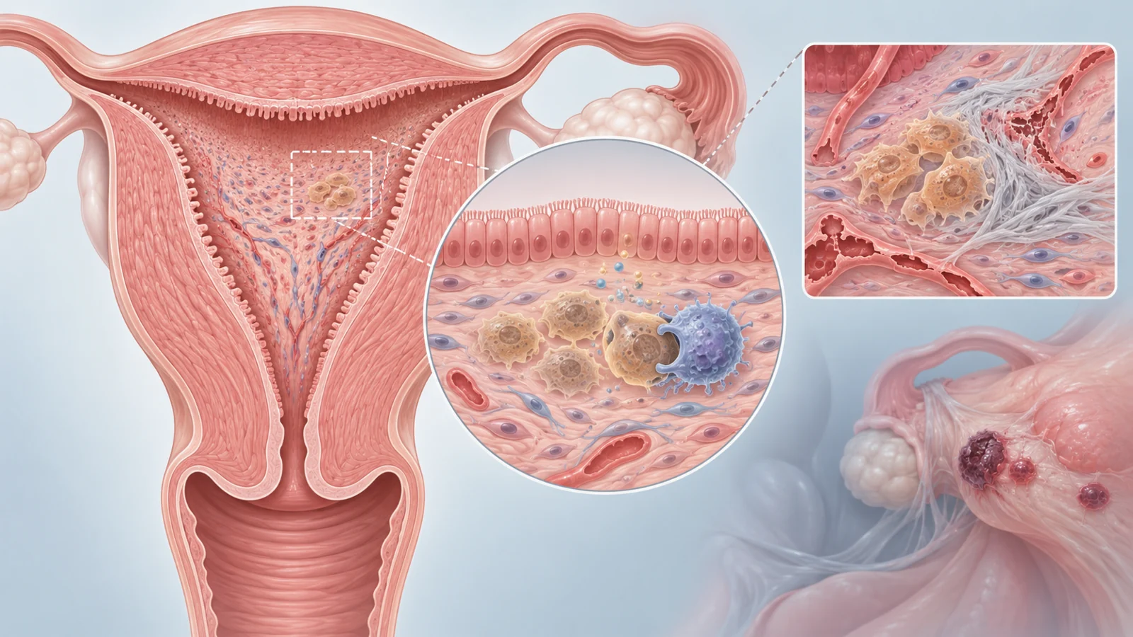
Cellular senescence is a state in which a cell permanently stops dividing while retaining active functions. Senescent cells can change shape and metabolism, respond to their surroundings and release signaling molecules known collectively as the senescence-associated secretory phenotype, or SASP. These secretions include inflammatory signals, chemokines and growth factors that can influence nearby cells and immune activity.
In laboratory cultures, senescent endometrial cells often become larger and flatter, with visible changes in the cytoplasm. Cells in living tissue may not look so different because their appearance is shaped by the surrounding architecture. Researchers therefore look for combinations of features such as increased SA-β-gal activity and higher expression of p16, also called CDKN2A, or p21, also called CDKN1A. None of these markers alone captures the whole biological state, a limitation that becomes important when findings from different studies are compared.
During the proliferative phase of the cycle, rising estrogen supports the expansion of stromal and perivascular cells. The review describes estrogen-related telomerase activity as one influence that can delay senescence while the lining grows. Later, progesterone becomes the dominant hormone and helps prepare the tissue for a possible embryo. Supporting endometrial stromal cells begin decidualization, a specialization process that reshapes the lining for implantation.
Some decidualized cells then enter an acute senescence-like state. Their secretions help create a measured early inflammatory response, promote decidual expansion, induce factors associated with implantation and recruit immune cells. Decidual natural killer cells can recognize and clear senescent-like decidual cells by releasing perforin and granzymes. The local tissue becomes more structurally permissive in selected regions, allowing the embryo to invade without turning the whole lining into an uncontrolled inflammatory environment.
This sequence explains the concern behind indiscriminate removal. Suppressing every senescence-like cell during early decidualization could leave the tissue too stable and insufficiently remodeled. Accumulation of uncleared cells could push the system in the opposite direction, with excessive SASP signaling, matrix disruption and reduced responsiveness to progesterone. The useful target is therefore not senescence in the abstract, but a misplaced, excessive or poorly cleared senescent population.
The same label describes different problems in different tissues
Endometriosis illustrates the inflammatory side of the story. In this disorder, tissue resembling endometrium grows outside the uterus and forms lesions in locations where it does not belong. The review summarizes findings of higher p16 expression in deep infiltrating lesions than in matched endometrium, along with increased IL-1β. Peritoneal fluid and tissue surrounding lesions also contain inflammatory cytokines such as IL-6, IL-8 and IL-1β, which overlap with components of the SASP. Endometrial stromal cells from people with endometriosis showed more SA-β-gal activity even without an external stimulus than cells from healthy controls in the cited studies.
Thin endometrium presents a different pattern. The review describes thin endometrium as a maximal proliferative-phase thickness below 7 millimeters, a finding associated with reduced receptivity and implantation difficulties. Single-cell analyses identified aberrant senescence as a feature of this condition. Senescence in endometrial stem or perivascular populations may limit proliferation and differentiation, reducing the tissue’s ability to regenerate. Epithelial cells show higher p16 and p21 expression, while perivascular cells display senescence-related gene activity alongside impaired support for vascular growth.
The immune environment may add to the problem. The review reports fewer uterine natural killer cells with senescent-cell clearance capacity in thin endometrium. Persistent cells can then continue producing SASP factors, while impaired vascular support and stromal changes create conditions associated with fibrosis. Here the concern is not primarily an ectopic inflammatory lesion, as in endometriosis, but a lining whose regenerative and vascular compartments may not rebuild adequately.
Intrauterine adhesions, also called Asherman syndrome, are different again. They are scars inside the uterine cavity that can form after damage to the basal endometrial layer, often following an intrauterine procedure. Because cyclic regeneration depends on a functioning microvascular network, injury can disrupt the capillary support needed for repair. The review describes increased p16 and p21 in endometrial endothelial cells from affected patients, together with reduced proliferation and impaired angiogenic capacity.
Senescent endothelial cells may fail to restore local blood-vessel support. The resulting tissue stress can activate stromal fibroblasts and promote scar deposition. In the studies summarized, stromal cells exposed to material from senescent endothelial cells increased fibrosis-related markers. PAI-1 produced by stromal cells was also described as a possible contributor to endothelial senescence through uPAR, with TGF-β amplifying PAI-1 production in an experimental feedback cycle. The cellular location matters: a signal associated with endometriotic inflammation is not the same problem as vascular failure inside scarred uterine tissue.
Implantation failure does not point in only one direction
The review also brings together findings from recurrent implantation failure and recurrent pregnancy loss, and they do not form a simple high-senescence versus low-senescence pattern. In some recurrent implantation failure samples, stromal cells undergoing decidualization in culture produced less IL-6 and showed lower levels of the decidual senescence marker DIO2 than control cells. PAI-1 and the proportion of p16-positive endometrial cells were also reported as lower in particular patient comparisons, including comparison with people who had successful pregnancies. These observations fit a possibility in which too little of the normal decidual senescence program impairs receptivity.
Other experiments in cells from patients with impaired receptivity showed premature senescence during the proliferative phase, including higher SA-β-gal activity and CDKN2A expression. Reduced CDC42 in patient-derived stromal cells was linked experimentally to Wnt activation, higher p21 and impaired decidualization. These are different tissue and laboratory contexts from the reduced signals described during decidualization, so they should not be collapsed into one direction of change.
Recurrent pregnancy loss adds another distinction. Mid-luteal endometrial tissue from affected patients showed what the review calls a pro-senescent decidual response, with increased markers including DIO2 and SCARA5. Decidual natural killer cells normally help maintain tissue balance by clearing senescent-like decidual cells. In recurrent pregnancy loss, insufficient clearance or altered recognition may allow excess cells to remain during decidualization, reducing receptivity. Animal work summarized in the review also connected a high-leucine diet with decidual senescence and embryonic loss, while a TNFSF14-related natural-killer-cell pathway counteracted leucine-induced senescence in mice.
These findings demonstrate why the timing, cell identity and experimental setting matter. A low signal during one decidualization comparison, premature senescence in another stromal-cell experiment and excess uncleared senescence in recurrent pregnancy loss may all be relevant without describing the same biological event. The review highlights the pattern but does not resolve the differences into a single diagnostic rule.
Treatment may need to change the signal rather than erase the cell
The therapeutic ideas discussed in the review fall into several broad categories. Senolytics aim to eliminate selected senescent cells. Other approaches seek to reduce the inflammatory secretions they produce or alter regulatory pathways that sustain the state. This distinction is crucial in the endometrium because removing cells could also remove a temporary function needed for decidualization and implantation.
Quercetin is the clearest concrete example in the review’s discussion. In animal models of endometriosis, quercetin reduced lesion size and improved tissue remodeling. That result supports further investigation of senolytic strategies in a disease where inflammatory senescence-like features are present, but it is not evidence that quercetin improves implantation or pregnancy outcomes in humans. The review also discusses metformin as a metabolic and inflammatory modulator and rapamycin as an mTOR inhibitor. These examples are presented as ways to influence the senescence-inflammation cycle, not as established endometrial treatments.
For intrauterine adhesions, inhibiting PAI-1 or targeting senescent endothelial cells is discussed as a possible way to interrupt the interaction between vascular dysfunction and fibrosis. For thin endometrium, the review draws a proposed comparison with chronic wounds, where excess senescent fibroblasts and weak immune clearance can impede repair and where senolytic treatment has promoted wound healing. That analogy suggests a testable direction for endometrial research, but wound-healing results do not demonstrate efficacy in thin endometrium.
The most difficult design problem is selectivity. A useful intervention would need to recognize pathological accumulation or harmful secretory activity while preserving the short-lived senescence-like changes that help prepare the lining for an embryo. The supplied evidence does not establish a treatment schedule that achieves this balance.
What the current evidence can and cannot distinguish
The research summarized by the review spans several levels of evidence. A tissue sample may show a senescence-associated signature through p16, p21, SA-β-gal or SASP-related molecules. A cultured-cell experiment may perturb a pathway and observe changes in proliferation, decidualization or inflammatory signaling. An animal experiment may test whether an intervention changes lesion size or tissue behavior. These are informative steps, but they answer different questions.
A marker pattern does not by itself show that senescence is the sole cause of a disorder. A result in patient-derived cells does not automatically predict what will happen in a living uterus, and a change in an animal model does not establish a useful reproductive treatment in humans. The review itself identifies small sample sizes, substantial reliance on in vitro systems, limited in vivo or translational validation and a lack of standardized senescence markers for the endometrium. Those limitations make cross-study comparisons difficult, especially when one study is measuring normal decidualization and another is examining disease-associated tissue damage.
The field therefore needs more than a larger list of senescence markers. It needs ways to distinguish cell type, anatomical location, timing within the cycle, secretory behavior and clearance status, followed by evidence that a targeted intervention preserves implantation-related function. A validated threshold separating beneficial remodeling from pathological accumulation had not been established by the material considered here. Reproductive outcomes in humans would be the decisive test of whether a proposed treatment helps the people it is meant to serve.
What selective treatment could mean for reproductive health
The possibility raised by this 2025 review is not the blanket removal of senescent cells. It is a more precise form of tissue repair: restoring a damaged lining’s ability to regenerate, support blood-vessel function and prepare for implantation while leaving normal decidual remodeling intact. Such treatment could eventually benefit people whose reproductive problems involve defects in endometrial regeneration, vascular support, scarring or the clearance of senescent-like cells.
That benefit remains conditional. Researchers would first need to distinguish pathological senescence from the temporary cell changes that help organize implantation, then show that an intervention produces safe and meaningful reproductive outcomes in humans. The review makes the reason for that standard clear: in the womb lining, the same broad cellular program can participate in both repair and disease, depending on its timing, location and control.
Key Sources
- Primary source: Cellular Senescence in Endometrium: A Pivotal Regulator in Physiological Remodeling and Pathological Disorders.
DOI or Journal Page · Review or Meta-Analysis · 2025-10-20
Keep exploring