
Research Signal – Source date 2025-09-02
Creatine helps cells manage energy, and getting it into cells depends in part on a membrane protein called the human creatine transporter. When this transporter is impaired, inherited SLC6A8 mutations can cause cerebral creatine deficiency syndrome 1, a neurological disorder associated with intellectual disability, speech delay, and seizures. In some cancers, prior research has motivated a different idea: blocking the transporter might limit a tumor’s access to creatine.
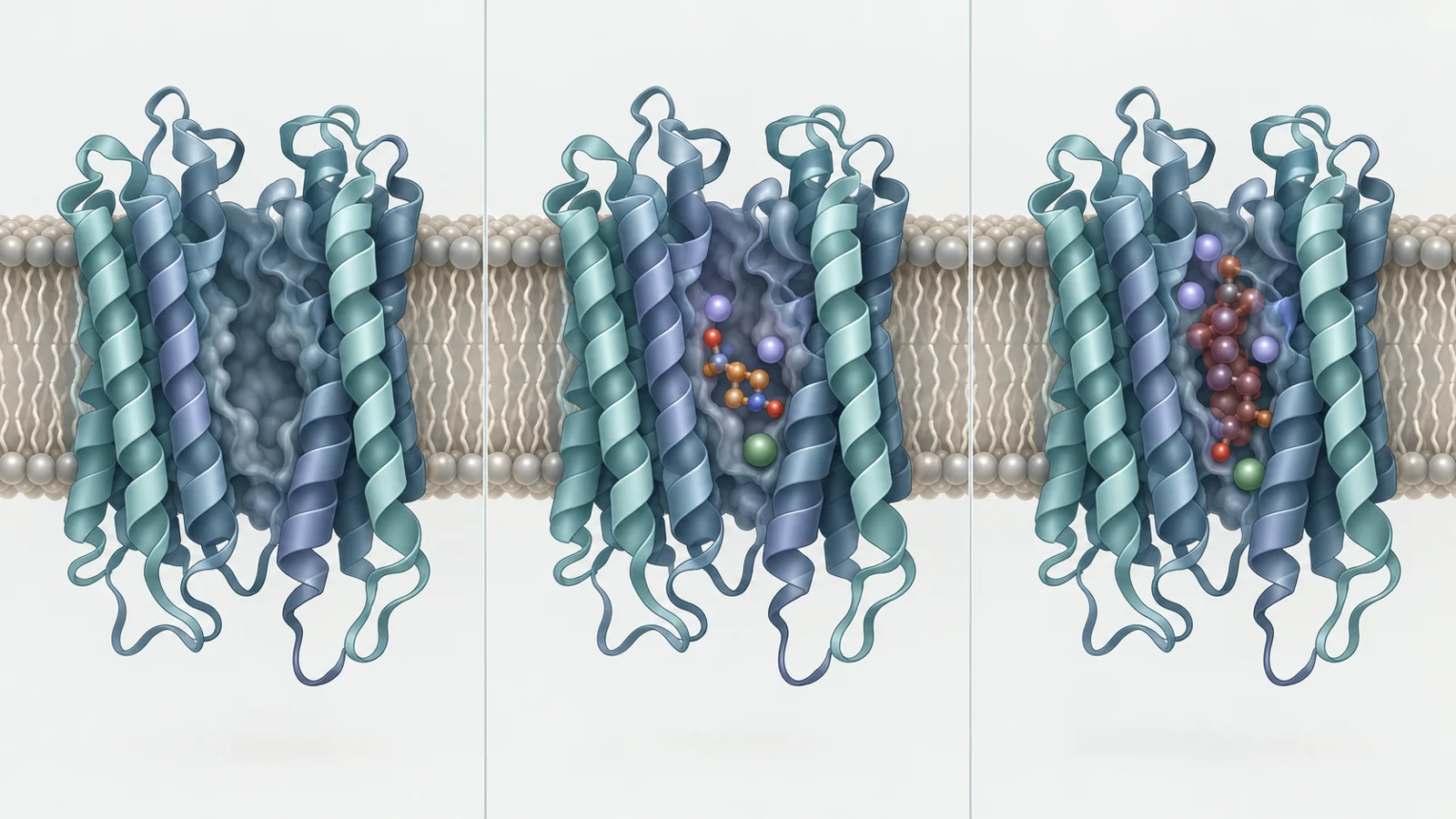
A study published on September 2, 2025, offers a detailed view of the transporter’s binding pocket. Researchers used cryo-electron microscopy to resolve purified human transporter in three conditions: empty, bound to creatine, and bound to RGX202, a competing inhibitor. Binding measurements and tests of altered pocket residues add support for how these molecules interact. Together, the work explains a molecular contest at the transporter. It does not show that blocking it benefits patients or that a disease-associated variant can be corrected.
Three snapshots of one transporter
The team produced recombinant human creatine transporter, or hCRT, and determined three cryo-EM structures. The empty, creatine-bound, and RGX202-bound structures were resolved at 3.28, 2.85, and 2.75 angstroms, respectively. These are snapshots of purified protein under different conditions, not a recording of creatine moving through a cell.
The transporter has 12 membrane-spanning helices arranged in a fold shared with other members of the neurotransmitter sodium symporter family. That shared architecture does not mean the family members carry the same cargo. The dopamine and serotonin transporters, for example, handle different molecules. The hCRT structures show how a related overall design can accommodate creatine through a more specialized pocket.
Creatine was held in a central pocket between parts of several membrane-spanning helices. Its position was oriented roughly along the plane of the membrane, and the bound structure was occluded: the molecule was enclosed, with neither an open route to the outside nor one to the cell interior. A bend in helix TM1a closed the inner route. A hydrophobic cap formed in part by Y148 and F315 sat over the binding site.
The transporter also relies on sodium and chloride ions. In the creatine-bound structure, researchers observed two sodium ions and one chloride ion in positions coordinated by transporter residues. One sodium site lies near the creatine carboxyl group; the other sodium and the chloride sit among nearby helices. These observed ion positions help describe the binding environment, although a structure alone does not show the full sequence of ion and substrate movements during transport.
A pocket tuned to creatine
The pocket’s local chemistry helps explain why hCRT recognizes creatine. The molecule’s guanidinium group sits among aromatic residues F68, Y148, and F315. The researchers describe potential hydrogen bonds involving Y148, backbone atoms, and water, alongside interactions between the aromatic rings and creatine. A nearby cysteine, C144, provides another contact with the guanidinium group.
Experiments that replaced selected residues support the importance of these contacts. Replacing aromatic residues with alanine reduced creatine affinity substantially; replacing C144 with alanine reduced it by more than 30-fold. These results connect particular parts of the pocket to binding in the tested system. They do not, by themselves, specify how each residue contributes to transport in a living tissue.
Comparison with related transporters highlighted a distinctive combination of residues around the pocket. In particular, C144 together with F68 and G71 helps shape the local environment around creatine’s guanidinium group. That combination offers a structural explanation for cargo selectivity: a shared transporter framework can be adapted by a small set of local differences to recognize a different molecule.
The inner gate shifts between observed states
When the researchers compared the empty and creatine-bound structures, the largest change they described involved TM1a, a helix that helps gate the inward-facing side. It tilted by about 21 degrees between the observed states. The movement is consistent with an inner gate opening to let bound creatine go toward the cell interior. A smaller shift in TM6b and changes in nearby residues accompany that rearrangement.
The comparison also showed changes around the ion sites. In the empty structure, the two sodium ions were not resolved, while the chloride remained visible. Residues near a sodium site had shifted, including a substantial rotation in S417, which helps coordinate that site in the creatine-bound structure. These differences suggest how ion coordination and gate position are linked, but they are comparisons between captured structures rather than direct observations of a transport cycle unfolding.
To fill in a state not captured by cryo-EM, the authors used AlphaFold2 to predict an outward-open conformation. In that model, the outer side of the transporter opens while the inner gate is closed, offering a plausible route for creatine entry before the transporter becomes occluded. The proposed sequence—entry, closure around the substrate, then opening toward the cell interior—is a mechanistic interpretation supported by the observed structures and a predicted state. The outward-open position itself was modeled, not experimentally resolved in this study.
RGX202 occupies creatine’s place
RGX202 binds in the same central pocket as creatine. Its guanidinium group and terminal carboxylic acid give it a chemical profile that resembles key parts of creatine, while a flexible carbon chain connects them. In the cryo-EM structure, RGX202 sits in a similar orientation and the transporter remains occluded. The overlap provides a direct structural explanation for competition: the inhibitor takes up space creatine would use.
Surface plasmon resonance measurements found similar affinities for the purified transporter: 3.2 micromolar for creatine and 3.39 micromolar for RGX202. The inhibitor’s bulkier chain extends closer to TM1 and may make additional contacts with backbone atoms in that region. The observed structure also showed a shift in one sodium ion’s position compared with the creatine-bound state. Mutating selected pocket residues reduced RGX202 binding affinity by roughly 30-fold, supporting their role in the interaction.
These findings explain how RGX202 can bind and compete at the molecular level. They do not establish how much transporter activity it blocks in a tumor, whether that changes tumor growth in people, or what benefits and risks treatment might have. The study’s structural and binding evidence addresses the first step in that chain, not the clinical outcome.
Disease variants may disrupt several parts of the job
The transporter’s role in the brain gives the structural work another connection to human disease. Mutations in SLC6A8 are linked to cerebral creatine deficiency syndrome 1. The researchers mapped selected disease-associated variants onto their structures and measured protein expression for a panel of variants. Several were present at levels comparable to the reference transporter in the cell-based expression measurements.
Comparable expression offers a useful correction to a simple assumption: a dysfunctional transporter need not always be scarce. A protein can be present and still have trouble binding creatine, coordinating ions, moving between conformations, or maintaining its structure. The team grouped mapped variants by their locations and proposed possible effects on the ion and substrate pathway, folding, helix interfaces, or coupling between structural regions.
Those categories are structural interpretations, not a proven explanation for every variant in a patient. The expression blots address abundance in the tested cells; they do not show that each variant reaches the correct place in relevant tissue or transports creatine normally. Nor do the structures predict an individual’s symptoms or show that a particular intervention can restore uptake.
How a creatine transport map could guide future treatments
A clearer account of how hCRT recognizes creatine and how RGX202 occupies its pocket could help researchers design and compare molecules that alter transporter activity. If those molecular insights translate into effective control of hCRT in relevant tissues, they could eventually support new approaches to limiting creatine supply in tumors or restoring transport in inherited deficiency. Both possibilities depend on substantial steps beyond purified-protein structures and binding tests, including showing what happens in cells, tissues, and clinical settings.
Key Sources
- Primary source: Structural insights into the substrate uptake and inhibition of the human creatine transporter (hCRT).
DOI or Journal Page · Peer-Reviewed Paper · 2025-09-02
Keep exploring