
Research Signal – Source date 2026-05-06
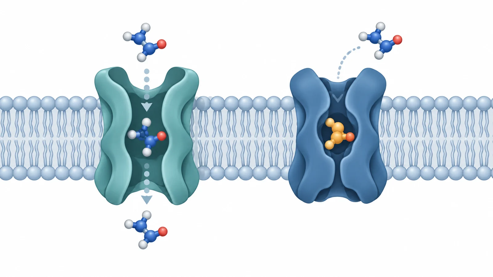
Creatine helps cells manage energy, and the protein that moves creatine into cells is called creatine transporter 1, or CT1. A molecule that reduces creatine uptake might seem to block this transporter. But the same readout can arise when CT1 carries a look-alike molecule through the cell membrane instead.
That difference matters when researchers search for compounds that could help explain how CT1 works or serve as starting points for future drug design. In a study published May 6, 2026, researchers used computer-built models of CT1 to screen for candidate molecules, then tested 16 of the highest-ranked compounds in laboratory assays. The experiments showed that a promising uptake-reducing hit could be transported cargo, while tiagabine and a related compound reduced uptake without detectable transporter-mediated currents in the reported cell tests.
A search built around CT1’s changing shapes
CT1, also known as SLC6A8, moves creatine across cell membranes and helps regulate cellular energy. Inherited changes that disrupt CT1 cause severe neurological disorders. CT1 overexpression has also been associated with cancer progression. Those findings give scientists reasons to study the transporter, but they do not show that inhibiting it would treat either condition.
When this project began, structural information for CT1 was limited. A transporter does not keep one fixed shape as it works, so a search based on a single model could miss features exposed in other states. The researchers built homology models—computer estimates based on related proteins—to represent CT1 in several conformations, including outward-open, occluded, and inward-open states.
They drew on multiple related-protein structures and evaluated the resulting models with computational quality checks. Those models gave the virtual screen different views of the transporter’s potential binding site. The computer search ranked compounds for follow-up; ranking was a way to select candidates for testing, not evidence by itself that any compound would bind CT1 or change its activity in cells.
The uptake result did not reveal the whole story
The team tested 16 top-ranked compounds in vitro. Compound 11 reduced creatine uptake with an inhibitory concentration, or IC₅₀, comparable to that of ompenaclid, a reference ligand used in the study. In plain terms, a similar concentration range was needed to produce the measured inhibition under the reported assay conditions. That made compound 11 an interesting hit, but an uptake measurement alone could not show whether CT1 had been blocked or had transported the compound instead.
To investigate, the researchers recorded currents from cells expressing CT1 while applying the candidate compounds. Currents are a measurable electrical response associated with transporter activity in this assay. Compounds 1 and 2 produced responses that the authors interpreted as evidence that CT1 transported them as alternative substrates. Similar inward currents were observed with compounds 8, 11, and 12, also supporting their classification as transported ligands.
Tiagabine, numbered compound 13 in the paper, and its analogue 16 produced a different pattern. They did not evoke detectable transporter-mediated currents in the reported recordings. Both also reduced creatine uptake, supporting their classification as nontransported inhibitors in these assays. GlyT1 inhibitors 14 and 15 also did not evoke detectable currents, though the reported uptake-inhibitor finding highlighted tiagabine and analogue 16.
The distinction is specific to the experiments described: the uptake result showed less creatine transport, while the current recordings helped the researchers assess whether a tested molecule was itself transported. The two measurements answer different questions. That is why compound 11’s comparable IC₅₀ made it a useful lead to examine, but did not make it equivalent to tiagabine or analogue 16 as a nontransported inhibitor.
Size may help turn a ligand into a blocker
The researchers used docking and molecular-dynamics simulations to examine how compounds might fit into CT1. In their simulations, compound 11 occupied the primary binding site with a stable pose. Its carboxyl group formed modeled hydrogen-bond interactions with Gly73 and Tyr148 and coordinated a sodium ion, resembling interactions attributed to the carboxyl group of ompenaclid.
These are computationally modeled interactions, not direct observations of compound 11 bound to CT1. The authors proposed that a carboxylate group paired with a protonatable basic center—such as an imidazole ring or piperidine—could help compounds interact with the transporter. They also suggested that greater size and lipophilicity may favor blocking over transport. In their interpretation, compound 11’s relatively compact structure may help explain why it can be carried through CT1 despite inhibiting creatine uptake.
That proposal points to a design idea rather than a demonstrated recipe: researchers could explore expanding compound 11’s imidazole-based scaffold to make bulkier derivatives, then test whether those changes shift the balance toward inhibition without transport. The screen and assays identify a starting point for such work; they do not establish that a larger derivative would bind as predicted or make a better inhibitor.
A later structural check supported the modeling approach
After the study work was completed, cryo-electron microscopy structures of human CT1 became available. The authors then compared those experimental structures with their earlier homology models. They reported overall agreement, including alignment of key features around the binding site.
This retrospective comparison supports the idea that the models captured useful aspects of CT1’s structure and could serve as a basis for screening. It is a check on the protein models, not a confirmation of every simulated ligand pose. Nor does agreement in this study show that the same screening approach will work equally well for other chemical families or targets.
Early leads still need a longer path
The evidence here comes from computational modeling, simulations, and cell-based experiments. It supports identifying compounds that affect creatine uptake and distinguishing candidates that appeared to be transported from those that inhibited uptake without detectable currents in the reported assays. It does not establish what these compounds would do in animals or people, whether they are safe, or whether changing CT1 activity would benefit people with a particular disease.
The disease context remains a reason for scientific interest, not a treatment claim. The researchers’ results are useful at an earlier stage: they offer candidate molecules and a way to connect structural hypotheses with laboratory measurements. Further evidence would be needed to determine how reliably the proposed interactions explain the observed activity and what biological consequences CT1 inhibition might have beyond these cell tests.
What better creatine transporter blockers could enable
If future work confirms that compounds can be designed to inhibit CT1 without being transported, researchers could gain more precise tools for studying how creatine uptake affects cellular energy and for testing whether CT1 is a useful target in disease research. The path to any practical or clinical use depends on resolving the remaining questions about binding, selectivity, safety, and effects in living systems; this study supplies early laboratory leads, not that outcome.
Key Sources
- Primary source: Structure-Based Search for Novel Creatine Transporter Inhibitors.
DOI or Journal Page · Peer-Reviewed Paper · 2026-05-06
Keep exploring