
Research Signal – Source date 2024-03-30
The puzzle behind an apparently normal scan
A person can have serious language difficulties even when a conventional brain scan does not show an obvious structural abnormality. A March 30, 2024 investigation explored that puzzle in two people with creatine transporter deficiency, an inherited disorder that prevents creatine from being transported into the brain and cells.
Both patients had unremarkable conventional MRI findings. Their diagnoses were supported by a known defective creatine transporter gene and by magnetic resonance spectroscopy showing minimal brain creatine. Yet both also had language impairment, with different degrees of severity. The study asked whether a more specialized form of MRI could reveal a pathway-level difference that a standard scan did not make visible.
Two patients with different language difficulties
The two patients were 28 and 18 years old and both were right-handed. The older patient had intellectual disability and substantial speech impairment, including severe difficulty retrieving words. His dependence was significant enough that he later required institutional care after the death of his grandmother.
The younger patient also had intellectual disability, but it was milder: he was able to attend school with sufficient proficiency, and his verbal impairment was described as mild. That contrast gave the researchers a limited but important comparison. The disorder was the same, while the language difficulties were not identical.
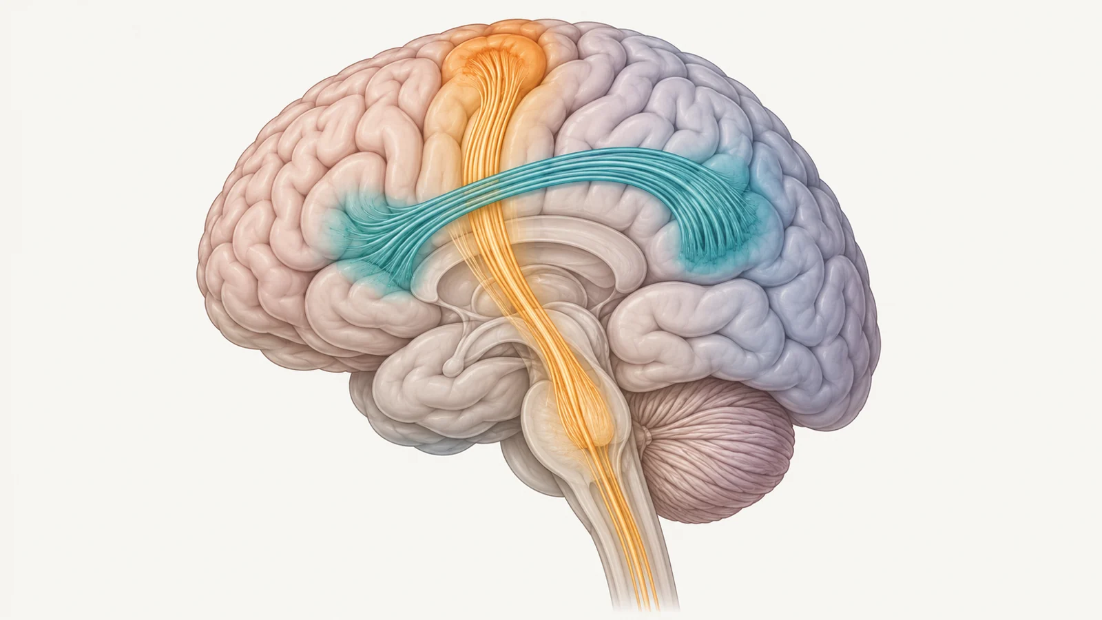
Creatine has an important role in brain energy metabolism, but the study focused on a less explored question: whether a lack of brain creatine might be associated with observable differences in the white-matter connections that support language. The researchers concentrated on the left arcuate fasciculus, a bundle linking language-associated regions commonly known as Wernicke’s and Broca’s areas. The pathway is relevant to speech, word retrieval, repetition and comprehension.
What tractography added
The researchers used diffusion MRI tractography to examine the structure of the two patients’ white-matter pathways. Tractography estimates the course of fiber bundles, while fractional anisotropy, or FA, provides a diffusion-derived measurement used here to compare the directional organization of those bundles.
The mean FA of the left arcuate fasciculus was lower in both patients than in the healthy-control measurements. The authors reported that the difference was statistically significant despite the small number of patients. The patient with the greater language impairment had the lower FA value, while the patient with milder verbal impairment had the higher value. Across these two observations, the language difficulty and the tract measurement showed a descriptive correspondence.
The researchers also examined the corticospinal, or pyramidal, tract, a pathway associated with motor control. Its FA values were comparable to those of the healthy controls in both patients. This comparison mattered because it suggested that the measured difference was not simply an obvious alteration across every white-matter pathway that was examined.
The result does not mean that the left arcuate fasciculus is the only affected pathway in the brain. The study compared one language-relevant tract with one motor tract, not every white-matter connection. Its contribution was narrower: it identified a selective difference in a pathway with a plausible relationship to the patients’ language difficulties, even though conventional MRI appeared unremarkable.
The finding has more than one possible explanation
The scans show an association, but they do not reveal the direction of the developmental process. One possibility is that the lack of brain creatine directly affected the development of the arcuate fasciculus, perhaps through effects on the biology that supports white-matter maturation. Another is that impaired development or reduced use of the language areas connected by the tract led to its underdevelopment. In that account, the pathway difference could be partly a consequence of altered language development rather than its initial cause.
The authors also discuss the possibility that creatine deficiency affects cortical cells and synaptic structures, which could influence the language regions and the connections that arise from them. These possibilities remain explanations to investigate, not conclusions established by the images.
The two adult scans cannot determine when the pathway difference emerged. They also cannot show whether the lower FA caused the language difficulties, resulted from them, or reflected an interaction between both processes. The statistical result indicates that the measurements differed from the comparison data in this study; it does not by itself establish a clinical threshold, predict an individual’s language ability or show that the same pattern applies throughout creatine transporter deficiency.
That distinction is especially important because the study involved only two diagnosed patients. Their different levels of language impairment make the comparison informative, but the correspondence between one patient’s lower FA and greater difficulty is descriptive rather than a validated clinical relationship. The findings point toward a possible region-specific effect without resolving how common it is or how it develops.
What this could change for understanding language disability
Connecting creatine transporter deficiency with specific changes in language-relevant pathways could eventually give researchers a more precise biological explanation for why language development is affected. If larger studies reproduce the pattern and clarify whether pathway development contributes to language disability, follows it, or both, clinicians may also gain a clearer way to explain differing language difficulties to affected families.
That potential benefit is conditional and explanatory. This study did not show that the pathway can be used to diagnose an individual, forecast recovery or select a treatment. Its value at the time of publication was to make a hidden anatomical question more precise: when standard MRI looks ordinary, examining the organization of a particular language pathway may reveal a difference worth investigating.
Key Sources
- Primary source: Selective Alteration of the Left Arcuate Fasciculus in Two Patients Affected by Creatine Transporter Deficiency.
DOI or Journal Page · Peer-Reviewed Paper · 2024-03-30
Keep exploring