
Research Signal – Source date 2024-06-13
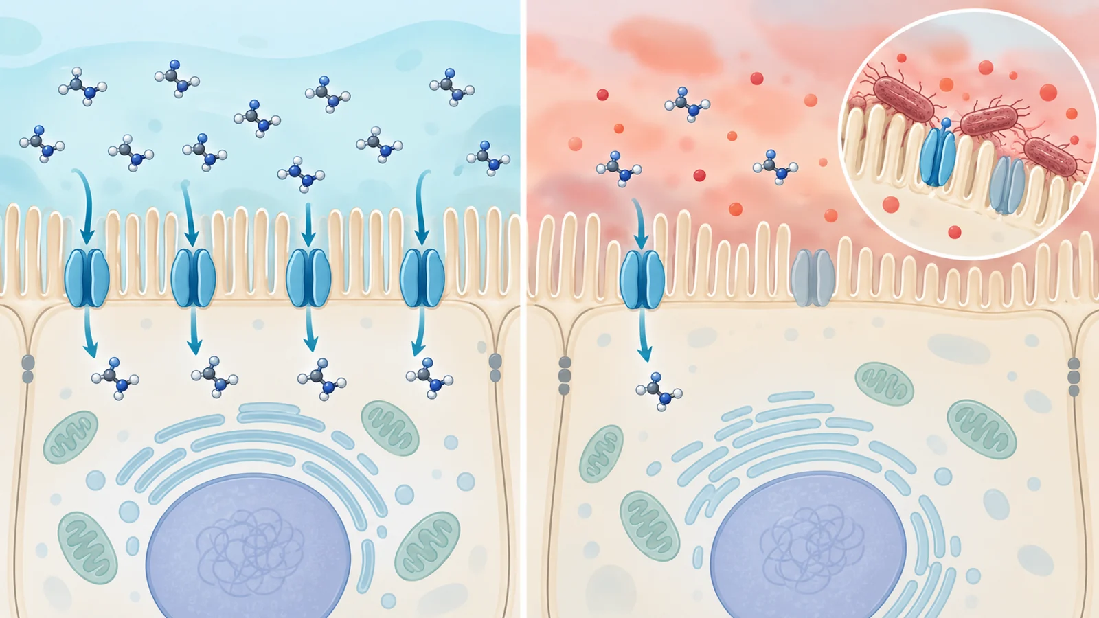
Creatine is often discussed as a nutrient available from food, but availability outside a cell is only part of the story. Intestinal epithelial cells must also move creatine across their surface and into the cell before they can use it in their energy system. A study published on June 13, 2024, examined whether inflammation and an adherent-invasive strain of Escherichia coli affect that entry process.
The research combined cultured intestinal cells, intestinal tissue from rodents, mouse-derived organoids, and healthy-human biopsy samples. It found lower activity and expression of the creatine transporter CrT1 in several inflammatory settings. The findings make intestinal creatine handling a plausible part of the biology of inflammatory bowel disease, but they do not show that transporter changes cause the disease or that creatine treatment improves it.
The question is about cellular access
Creatine is a non-protein amino acid involved in the creatine–phosphocreatine system. Creatine kinase helps convert creatine and ATP into phosphocreatine and ADP, allowing cells to store and distribute high-energy phosphate groups between sites where energy is produced and sites where it is needed. Intestinal epithelial cells are among the cells that carry out energy-demanding work while maintaining the barrier and absorbing nutrients.
The transporter examined in this study is CrT1, encoded by the SLC6A8 gene. It is a membrane protein that helps bring creatine into cells. The paper first asked where this transporter is found along the intestine, then tested what happened to its function and expression when intestinal cells were exposed to inflammatory signals or infected with a disease-associated bacterial strain.
In rat jejunum, imaging placed CrT1 on the brush border, the lumen-facing surface of absorptive cells. That location is consistent with a role in taking up material from the gut contents. Measurements in mouse and rat intestinal regions showed the highest CrT1 messenger RNA and protein levels in the jejunum. Samples from healthy human jejunal and colonic mucosa showed the same general regional pattern for protein, although this human comparison was narrower than the animal mapping.
Inflammatory cytokines reduced both transport and transporter production
The researchers used differentiated Caco-2 and IEC-6 cell monolayers as laboratory models of absorptive intestinal epithelial cells. They treated the cells with tumor necrosis factor alpha, or TNFα, and interleukin-1 beta, or IL1β. These cytokines are associated with intestinal inflammation.
After treatment, sodium-dependent creatine uptake fell in both cell lines. The uptake measurement used radiolabeled creatine applied to the apical, or lumen-facing, side of the cells. The reported assay used three samples per condition, written as n = 3; that count describes the uptake experiment and should not be generalized to every tissue, organoid, or expression comparison in the paper.
The researchers also measured the transporter itself. CrT1 mRNA and protein levels decreased after exposure to either cytokine in both cell models. These observations answer a different question from the uptake assay: they show that less transporter-related material was present, while the uptake result shows that the cells’ measured ability to take up creatine was reduced.
A promoter experiment provided another layer of evidence. In Caco-2 cells, the researchers introduced a 1,029-base-pair human CrT1 promoter and measured its activity after cytokine treatment. Activity declined after exposure to TNFα or IL1β. Because a promoter helps regulate transcription, this result supports a transcriptional contribution to the lower CrT1 expression. It does not identify the complete signaling pathway linking inflammatory exposure to the transporter, nor does it show that every reduction in uptake must arise from transcriptional regulation.
The pattern also appeared in a spontaneous ileitis model
The study then moved beyond cytokine-treated cell lines. SAMP1/YitFc mice develop spontaneous ileitis and are used as a model that resembles aspects of human inflammatory bowel disease, while AKR/J mice served as controls. CrT1 mRNA and protein levels were lower in the small-intestinal tissue of SAMP1 mice than in AKR controls.
This comparison matters because it asks whether reduced transporter expression is visible in intestinal tissue during naturally developing inflammation within the model, rather than only after researchers add a cytokine to cells in a dish. It still remains a model comparison, not a demonstration of the same process in people with Crohn’s disease or ulcerative colitis.
The researchers also grew three-dimensional small-intestinal organoids from SAMP1 and AKR tissue. CrT1 mRNA and protein were again lower in organoids from SAMP1 mice. Organoids preserve some features of intestinal tissue in a controlled setting, so they can help examine whether a tissue-associated expression pattern persists outside the original animal. They do not, by themselves, establish the amount of creatine entering the cells, the size of any energy deficit, or the clinical consequences of the expression change.
LF-82 added an infection-dependent wrinkle
The bacterial experiment used LF-82, an adherent-invasive E. coli strain associated with Crohn’s disease research. In cultured IEC-6 and Caco-2 monolayers, infection was followed by lower CrT1 mRNA. The result therefore extended the study from inflammatory cytokines to a tested microbial exposure, but the supplied results support an expression claim rather than a complete account of how infection changes creatine uptake or cellular energy handling.
The clearest context-dependent comparison came from organoid-derived monolayers. LF-82 reduced CrT1 mRNA in monolayers made from SAMP1 organoids. In monolayers made from AKR organoids, the change was not statistically significant. The authors interpreted this pattern as suggesting that bacterial effects on CrT1 may be more pronounced in intestinal tissue with pre-existing inflammation.
That interpretation does not establish why the two models responded differently. The difference could reflect features of the inflamed model, the tissue state retained by the organoids, or other model-specific factors. Nor does the result show that all E. coli strains affect the transporter in the same way. The study tested LF-82, and its bacterial expression findings should not be expanded into a general claim about intestinal infection.
What remains unresolved about inflammatory bowel disease
Across the experiments, the study connects several observations: CrT1 is concentrated in a region suited to nutrient absorption; cytokine exposure reduces measured creatine uptake and several markers of transporter expression; spontaneous ileitis is associated with lower CrT1 expression in mouse tissue and organoids; and LF-82 lowers CrT1 mRNA, with the strongest organoid-derived response observed in the SAMP1 inflammatory model.
The direction of causation remains open. CrT1 downregulation could contribute to reduced energy availability in intestinal epithelial cells, could result from chronic inflammation, or could participate in a feedback process involving both. The experiments do not establish that CrT1 dysregulation initiates inflammatory bowel disease in humans.
The healthy-human biopsies provide anatomical comparison material. They do not show how CrT1 responds in patients with inflammatory bowel disease, whether treatment changes transport, or whether altered transport predicts symptoms or disease activity. Likewise, lower transporter expression and lower uptake in experimental systems do not establish whole-body creatine deficiency or the magnitude of an intestinal energy deficit.
These boundaries also matter for supplementation. The study does not show that oral creatine restores CrT1 activity, overcomes reduced uptake, improves symptoms, induces remission, or is ineffective because transport is impaired. A dietary intervention would require evidence about transport, tissue biology, patient outcomes, dosing, and safety that these experiments did not provide.
What this could mean for treating intestinal inflammation
The potential clinical value lies in a future treatment capability: helping inflamed intestinal tissue maintain access to an energy-supporting nutrient while the underlying inflammation is controlled. Preserving or restoring creatine entry into intestinal epithelial cells could become an additional therapeutic approach if later work shows that impaired transport contributes meaningfully to disease and that correcting it safely improves outcomes that matter to patients.
For now, the study supplies an experimental starting point rather than a treatment. Its contribution is to show that intestinal creatine handling is regulated at the transporter level, can change under inflammatory and LF-82 exposure, and may be especially vulnerable in an already inflamed tissue model. Whether that biology can be modified in people, and whether modification would help them, remains to be established.
Key Sources
- Primary source: Intestinal Epithelial Creatine Transporter SLC6A8 Dysregulation in Inflammation and in Response to Adherent Invasive E. coli Infection.
DOI or Journal Page · Peer-Reviewed Paper · 2024-06-13
Keep exploring