
Research Signal – Source date 2021-08-12
Creatine accumulated in fetal sheep brains, but the clearest physiological difference appeared later in recovery from an oxygen shortage. In an experiment published on August 12, 2021, creatine-treated fetuses had lower arterial blood oxygen measurements during the second and third days after the challenge. Getting creatine into the brain and demonstrating protection against oxygen deprivation are separate questions; this study’s reported findings leave the second unresolved.
The timing makes the result particularly interesting. The researchers gave creatine for ten days before briefly interrupting the umbilical cord’s blood flow, then followed fetal physiology for another three days. The reported oxygen difference emerged well after the immediate challenge had ended.
Ten days of infusion before a ten-minute challenge
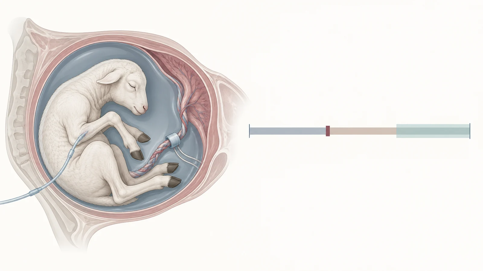
The experiment involved 28 pregnant ewes, surgically instrumented at 118 days of gestation. Starting at day 121, their fetuses received a constant intravenous infusion of either creatine or an equal volume of saline. Creatine was delivered directly to the fetus at 6 milligrams per kilogram per hour. This was a controlled fetal exposure, distinct from a pregnant mother taking an oral supplement.
After ten days, the researchers temporarily occluded the umbilical cord for ten minutes to produce what they described as mild, acute global hypoxia—an oxygen shortage affecting the fetus broadly. Other fetuses underwent a sham procedure. There were eight fetuses in the saline-plus-occlusion group, seven in the creatine-plus-occlusion group, six saline controls, and seven creatine controls.
That four-group design allowed the researchers to compare creatine with saline both with and without the oxygen challenge. The sham groups helped distinguish effects associated with creatine exposure from those associated with the cord occlusion. Cardiovascular measurements, arterial blood gases, blood metabolites, and plasma creatine were monitored before, during, and after the procedure.
Creatine in the blood and in the brain
The infusion increased plasma creatine concentrations fivefold. That measurement describes exposure in the circulating blood. The regional brain measurements answered a different question: whether creatine had accumulated in the tissues examined.
At the 72-hour endpoint after the challenge, increased total creatine was detected in cortical gray matter, the hippocampus, the thalamus, and the striatum. These endpoint measurements locate tissue accumulation at the end of observation; they do not provide a continuous record of brain creatine throughout the oxygen shortage and recovery.
The striatum also showed an increase in total creatine associated with the cord occlusion itself. The authors suggested that increasing creatine availability there might be a fetal adaptation to oxygen deprivation. That interpretation raises a biological possibility beyond the infused dose, although an adaptive role remains a hypothesis.
The oxygen difference appeared during later recovery
Before occlusion, the creatine infusion had no significant effect on the measurements reported in the abstract. The abstract also reports no alteration in fetal physiology during the occlusion or during early recovery, through the first 24 hours.
During hours 24–72, however, creatine-treated fetuses showed significantly lower arterial oxygen pressure and oxygen saturation in the reported analysis. These are related but distinct measurements. Oxygen pressure concerns oxygen dissolved in arterial blood; saturation describes how fully the blood’s hemoglobin is carrying oxygen. Both describe blood oxygenation, rather than directly measuring brain injury.
The delayed timing broadens the observation beyond the ten-minute event. An assessment confined to the challenge and its immediate aftermath would miss the period in which this difference was reported. The abstract establishes its direction and statistical significance, but does not provide enough detail to assess the size of the oxygen changes.
Why lower blood oxygen leaves the outcome open
The oxygen measurements alone cannot distinguish increased tissue oxygen use from impaired oxygen delivery, or determine whether the overall effect was beneficial or harmful. The authors proposed that fetal creatine supplementation might alter oxygen movement following the challenge. Establishing that explanation would require evidence connecting the blood measurements to what tissues were receiving and using.
Likewise, increased brain creatine does not establish that injury was prevented or neurological function improved. The physiological findings summarized here cannot settle those outcomes. Their interpretation is also bounded by groups of six to eight animals, direct intravenous dosing, a brief mild challenge, and observation through 72 hours. Those conditions cannot establish human pregnancy safety, a maternal oral dose, or long-term developmental effects.
What this could change
If increasing fetal creatine can eventually be shown to reduce oxygen-related injury, and a safe, practical way to deliver it before birth becomes available, this approach could offer preventive protection for babies at risk of oxygen deprivation around birth. The potential benefit would come from preparing vulnerable tissues before an oxygen shortage occurs. This sheep experiment contributes a concrete starting point for that possibility by showing fetal brain creatine loading alongside a recovery response that extends beyond the immediate event.
Key Sources
- Primary source: The physiological effects of creatine supplementation in fetal sheep before, during, and after umbilical cord occlusion and global hypoxia.
DOI or Journal Page · Peer-Reviewed Paper · 2021-08-12
Keep exploring