
Research Signal – Source date 2024-12-16
Removing damaged cells sounds like a straightforward way to help an injured liver. But some cells that have stopped dividing still perform useful work. Eliminating them can have very different consequences depending on whether they process nutrients, produce scar material, or maintain the boundary between blood and liver tissue.
A review published on December 16, 2024, brings these differences together to examine cellular senescence—a state in which cells stop dividing but remain metabolically active. It connects findings across liver cell types and diseases, including experiments in mice and observations in human tissue. Its contribution is an explanation of why targeting senescence can produce sharply different results, rather than a new demonstration of treatment benefit in patients.
A cell can stop dividing and keep sending messages
Senescence involves a durable arrest of the cell cycle, the process through which a cell grows and divides. DNA damage, shortening of the protective ends of chromosomes, and malfunctioning mitochondria can help trigger it. Proteins such as p21 and p16 participate in the molecular brakes that prevent further division.
An arrested cell is still alive. Senescent cells can activate survival mechanisms that resist apoptosis, the controlled process of cell death. They also continue to release molecules into their surroundings. This output, called the senescence-associated secretory phenotype, or SASP, includes inflammatory signals, growth regulators, and enzymes that remodel the material around cells.
Those secretions explain how a change inside one cell can affect an area of tissue. They can recruit or alter immune cells, influence nearby cells, and reinforce the senescent state. Their composition varies with the cell type, the stress that initiated senescence, and the time elapsed. A single label therefore covers cells sending substantially different chemical messages.
The review describes one liver-specific example involving hepatocytes, the cells responsible for much of the organ’s metabolic work. A signal released by senescent cells, CCL2, can stimulate liver macrophages—immune cells—to produce TGF-beta. That molecule can then promote senescence in neighboring hepatocytes. This sequence gives researchers a mechanism for studying how an initially local problem might spread and interfere with regeneration.
Three liver jobs with different consequences
Hepatocytes process nutrients and fats and carry out many of the liver’s essential functions. Their ability to divide also contributes to regeneration. When damaged hepatocytes enter senescence, arresting their growth can restrain potentially cancerous proliferation. Persistent senescence, however, can limit the liver’s capacity to renew its working cells and sustain inflammatory signaling.
Fat metabolism provides a more specific connection to disease. The review links hepatocyte senescence markers with impaired mitochondrial fat breakdown in mouse models and small human cohorts. When cells process fatty acids less effectively, triglycerides can accumulate. This helps explain the interest in senescence in metabolic dysfunction-associated steatotic liver disease, or MASLD, and its inflammatory form, MASH.
Hepatic stellate cells have a different role. During chronic injury, activated stellate cells multiply and produce extracellular matrix, the structural material that accumulates as scar tissue. Excessive deposition produces fibrosis; continued scarring can progress to cirrhosis and impair liver function.
For these scar-producing cells, entering senescence can interrupt a damaging process. The review describes how senescence limits further matrix deposition by activated stellate cells. They can also release matrix metalloproteinases, enzymes that break down existing matrix. Experiments in which key senescence pathways are disabled show the other side of this mechanism: stellate cells continue multiplying and depositing excessive scar material.
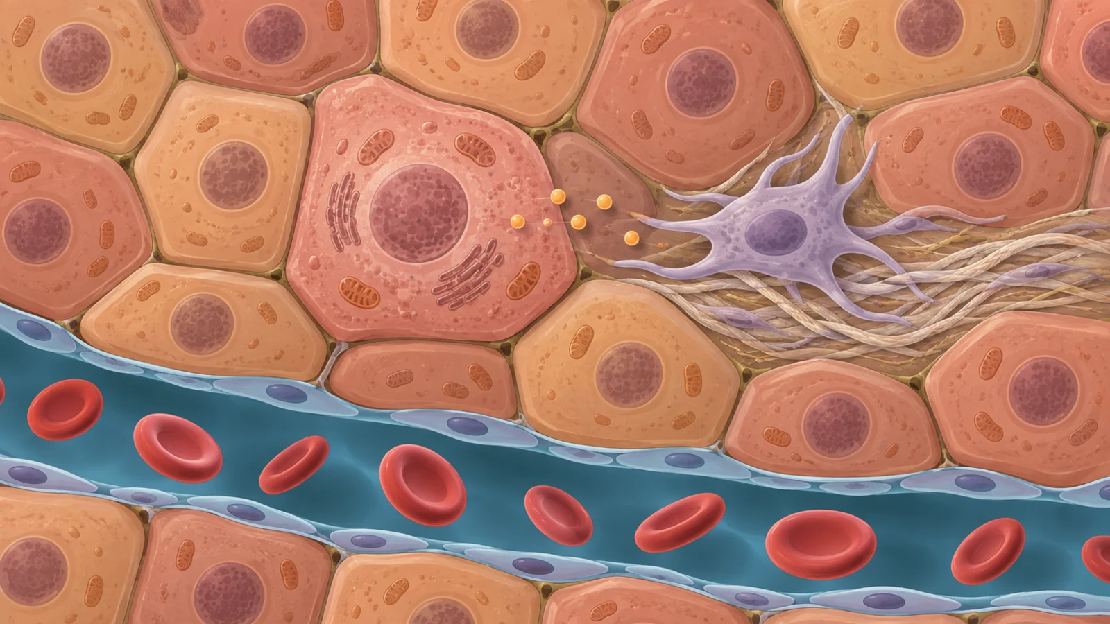
A third population forms the thin lining of the liver’s small blood channels, or sinusoids. These sinusoidal endothelial cells maintain the interface between circulating blood and liver tissue. The review reports that removing p16-positive lining cells from aged mouse livers disrupted that barrier and induced fibrosis around blood vessels. Here, eliminating cells selected by a senescence-associated marker created a structural problem in the tissue.
Together, these findings make cell identity central to the removal question. A hepatocyte with impaired metabolism, a stellate cell whose scar production has slowed, and a lining cell maintaining a blood–tissue boundary are not interchangeable targets. The same broad senescence label does not tell researchers what function will disappear when a cell is cleared.
What cell removal changed in mice
Senolytics are agents intended to eliminate senescent cells. The review discusses a combination of dasatinib and quercetin, often abbreviated D + Q, as one experimental approach. In older, obese, and diabetic mice, treatment with this combination reduced liver fat accumulation. Genetic removal of cells expressing p16 also reduced liver fat in the experiments described.
The measured outcome matters. Less fat in a mouse liver is a finding about steatosis, the buildup of fat within the organ. It helps support a functional connection between senescent cells and disturbed fat handling. It does not, by itself, establish recovery from advanced scarring or prevention of liver failure.
Engineered immune cells offer a different approach. Chimeric antigen receptor T cells, or CAR T cells, carry synthetic receptors that redirect their targeting. The review describes experimental CAR T cells designed to recognize senescent cells. In mice with liver fibrosis induced by prolonged exposure to carbon tetrachloride, these cells showed senolytic activity and improved fibrosis-related pathology.
That experiment addresses scar-related injury in a chemically induced model. The drug-combination experiments address fat accumulation in metabolic and aging settings. Reading the results separately preserves what each contributes: one tests whether removing selected cells can change liver fat, while the other tests an immune-cell strategy against experimentally induced fibrosis. Neither supplies a universal measure of how much “better” a liver becomes after senescent-cell removal.
Why an experimental target is not yet a patient treatment
Identifying the target is an unresolved practical difficulty. Researchers use several features to recognize senescence, including cell appearance, cell-cycle inhibitors such as p16 and p21, and activity of an enzyme called senescence-associated beta-galactosidase. These measurements provide clues, but their interpretation depends on the biological setting.
The review highlights limitations of beta-galactosidase testing. In laboratory cultures, crowded cells or cells deprived of serum can give misleading positive results. Some senescent cells may also lack the expected enzyme signal. Consequently, a change in one marker cannot automatically be read as a reliable count of harmful senescent cells. Selecting a treatment target requires evidence about both the cells’ state and their role in the tissue.
Human observations answer a different question from intervention experiments. Small patient cohorts and liver biopsies connect senescence markers with fat accumulation and greater MASLD or MASH severity. These associations make the biology relevant to human disease, but they cannot establish that senescence caused all of the observed damage or that clearing the marked cells would improve patients’ health. Chronic injury may itself help drive the senescent state.
Toxicity creates a separate obstacle even when a candidate reaches its intended target. Navitoclax, another agent discussed in the review, blocks proteins in the BCL-2 family that help cells survive. It has shown senolytic effects in experimental systems, including liver disease models, but its development is constrained by substantial blood-cell toxicities. These include thrombocytopenia, a reduction in platelets, and neutropenia, a reduction in a type of infection-fighting white blood cell. The review also identifies thrombocytopenia as a concern with dasatinib.
These are distinct gaps between an encouraging experiment and a usable therapy. Researchers need dependable ways to identify the relevant cells, evidence that intervening changes human disease, and a treatment whose harms remain acceptable. The review’s conclusion emphasizes the distance between preclinical findings and clinical translation. The evidence it brings together does not establish that senescent-cell clearance improves clinical outcomes in people with chronic liver disease or prevents cirrhosis, liver failure, or liver cancer.
What this could change
If the experimental findings translate into safe, durable human benefit, targeting senescence could provide another way to interrupt liver disease progression. Reducing fat-related injury could ease continuing stress on hepatocytes. Reducing fibrotic damage could help preserve the tissue structure on which liver function depends.
The downstream possibility is that patients could retain more functioning liver tissue for longer and progress more slowly toward cirrhosis. Reaching that point would turn changes observed in experimental liver fat and scarring into a clinically meaningful capability: slowing the loss of an organ’s ability to do its everyday work.
Key Sources
- Primary source: Navigating the complex role of senescence in liver disease.
DOI or Journal Page · Peer-Reviewed Paper · 2024-12-16
Keep exploring