
Research Signal – Source date 2022-01-27
A leukemia sample can contain a measurable immune checkpoint marker without giving doctors a clear answer about whether checkpoint-blocking treatment will help. The marker may offer useful information, but it is only one part of a changing interaction among leukemia cells, immune cells and the body’s response to treatment.
That is the central challenge in a review published on January 27, 2022, comparing laboratory approaches for following checkpoint blockade therapies in leukemia. The authors describe tests that might help assess treatment response and harm, but the review does not identify a validated leukemia-specific test that reliably picks out patients who will benefit.
Releasing immune restraints can have mixed effects
Immune checkpoint proteins act as restraints that help regulate immune activity. Cancer can use these pathways to dampen an attack by immune cells. Checkpoint blockade uses antibodies to interfere with those restraints, aiming to let immune cells act against cancer. The approach has benefited some people with blood cancers, but its effects in leukemia are more limited and uneven.
Treatment can also disturb the immune balance that checkpoints help maintain. The resulting immune-related adverse events can involve the skin, digestive system, liver, eyes, lungs or hormone-producing organs. Some complications may be reversible, while some endocrine effects can leave lasting organ dysfunction. The review notes that these adverse events have generally been less frequent with PD-1 or PD-L1 blockade than with CTLA-4 blockade, but that comparison should not be read as a leukemia-specific rate.
Resistance adds another reason to follow patients over time. Some people do not respond from the outset. In others, resistance may emerge as the disease adapts, or develop after a period of temporary disease control. A pretreatment sample therefore cannot describe every change that may matter during therapy.
A positive PD-L1 stain is only one clue
PD-L1 is a checkpoint protein that can be measured on cells. In some solid tumors, its expression helps guide treatment decisions. In leukemia, the evidence is less settled: responses have been observed even when tumor samples show low or no PD-L1, and levels may have different meanings across leukemia subtypes. The review cites a study linking higher PD-L1 expression with worse outcomes in a particular leukemia setting, but that association does not make PD-L1 a dependable stand-alone predictor of treatment benefit.
The test itself can also vary. Results depend on the antibody clone, staining method and threshold used to call a sample positive. Leukemia samples add complexity because they contain mixed cell populations in blood or bone marrow. Scoring systems developed for solid tumors count tumor cells, or tumor and other immune cells, in ways that cannot simply be transferred to leukemia.
Other tests ask different questions. Tumor mutational burden estimates the number of mutations in tumor DNA; more mutations may produce targets the immune system can recognize. A threshold of 10 mutations per megabase is used as a predictive factor in some contexts, yet the review describes conflicting evidence about how well this measure predicts response to checkpoint therapy. Microsatellite instability, a pattern of DNA changes used in some solid-tumor treatment decisions, also has uncertain relevance in blood cancers. One study cited by the review reported low microsatellite instability in chronic lymphocytic leukemia and none in the acute myeloid leukemia cases it examined.
These tests measure different features: a checkpoint protein, the number of mutations, or a pattern of DNA instability. None alone captures how leukemia cells and immune cells are interacting in a particular person, and evidence from other cancers cannot settle that question for leukemia.
Looking beyond the malignant cells
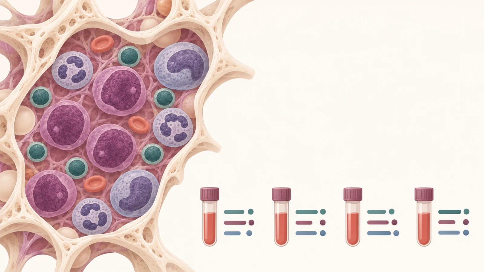
Routine leukemia diagnosis already combines several ways to characterize malignant cells, including their appearance, surface markers, chromosomes and molecular features. The review proposes adding more detailed profiling of immune cells in the bone marrow during follow-up, alongside those established assessments. Bone marrow is accessible for sampling, but it is also a varied environment containing leukemia cells, immune cells, supporting cells and other structures. Interpreting it calls for specialist expertise.
One suggested approach is a two-tube flow-cytometry panel designed for 13-color instruments. Flow cytometry labels cells so a laboratory can distinguish populations by their features. The proposed panel could help identify types of T cells, including cells involved in killing or coordinating immune responses, and measure checkpoint proteins and signs of activation. It could also help describe whether immune-cell populations change during treatment.
That is a proposed way to study the immune response, not a proven treatment-selection tool. The review does not show that using the panel improves outcomes, and different laboratories may not produce comparable results without agreed methods and further evaluation.
Following inflammation as well as response
Laboratory follow-up has a second purpose: noticing possible harm as immune activity changes. Common measurements such as blood-cell counts, C-reactive protein and interleukin-6 can describe aspects of inflammation. The review also points to a bone-marrow immune-cell population with low or absent HLA-DR, a marker on certain monocytes, as a possible area for study in leukemia patients receiving checkpoint therapy.
These measurements do not amount to a validated warning system for serious complications such as inflammation of the heart, brain or pituitary gland. The review calls for better ways to predict such events; it does not establish that serial inflammatory tests can tell which individual patient will develop them. The goal is to understand both disease response and treatment-related inflammation, while recognizing that the same laboratory result may not answer both questions.
Making results comparable across laboratories
A measurement becomes useful in routine care only if laboratories can produce and interpret it consistently. For checkpoint-marker staining, antibody clones, preparation and scoring thresholds can differ. Bone marrow and blood also contain mixtures of cells, which can complicate comparisons. Detailed immune profiling may require specialized skills and equipment that are not available in every hospital laboratory.
The review describes proposed methods and research findings rather than a single agreed follow-up algorithm. It calls for more standardization and evaluation, including suitable reagents and testing approaches that laboratories can adopt. Until methods are more consistent and tested in leukemia, a promising result from one study or laboratory may not translate into the same meaning elsewhere.
How this could shape the future
A more informative future visit could bring several kinds of evidence together: how the leukemia cells are changing, whether immune-cell populations are shifting, and whether blood or marrow tests show signs of harmful inflammation. For a person receiving checkpoint therapy and their care team, seeing those patterns across visits could support more grounded treatment discussions than relying on one checkpoint-marker result. That possibility depends on standardized assays and prospective validation showing that the measurements are reliable and clinically meaningful.
One nearer milestone would be a multicenter leukemia study demonstrating that the same defined set of measurements can be reproduced across laboratories and relates to both treatment response and adverse-event outcomes. The review provides a rationale for examining those pieces together; it does not establish that such a combined approach is ready to guide care.
Key Sources
- Primary source: Comparison of Laboratory Methods for the Clinical Follow Up of Checkpoint Blockade Therapies in Leukemia: Current Status and Challenges Ahead.
DOI or Journal Page · Review or Meta-Analysis · 2022-01-27
Keep exploring