
Research Signal – Source date 2021-06-19
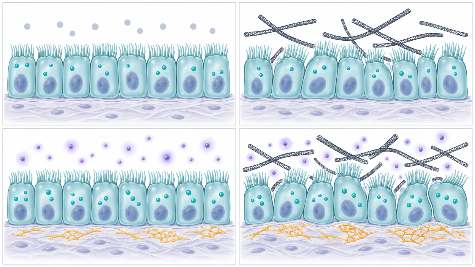
A material can alter how cells grow and communicate before it causes obvious cell death. A study published on June 19, 2021, examined that possibility for multi-walled carbon nanotubes, or MWCNTs: tiny tubular carbon structures with a fiber-like form. The experiment used cultured human bronchial epithelial cells called BEAS-2B, rather than a complete lung or a human population.
The researchers compared nanotubes alone with nanotubes given alongside transforming growth factor beta, usually abbreviated TGF-β. TGF-β is a signaling protein involved in fibrotic responses, including processes associated with lung scarring. The central question was whether a cell environment already receiving that signal would respond differently to the material.
The answer depended on which cellular behavior was measured. The nanotubes produced signs of oxidative stress and slower growth, while the combined treatment produced a broader set of senescence-associated and remodeling-related changes. The findings suggest an interaction worth studying, but they do not turn a culture experiment into evidence that exposed people will develop pulmonary fibrosis.
One exposure produced several different timelines
The study separated early stress, cell viability, proliferation and senescence-associated activity instead of treating them as one outcome. Reactive oxygen species, or ROS, increased about eight hours after nanotube exposure. ROS are chemically reactive molecules that can rise when cells are under oxidative stress, but an elevated signal is not the same as proof of irreversible damage.
After 24 hours, the viability assay detected no cytotoxicity at nanotube concentrations up to 50 micrograms per milliliter. In other words, the cells were not measurably killed under those assay conditions. A separate 48-hour proliferation assay showed a dose-dependent reduction in cell counts at concentrations above 0.5 micrograms per milliliter. Fewer cells after two days can reflect inhibited growth, so this result should not be collapsed into the short-term viability result.
At 72 hours, the researchers measured senescence-associated β-galactosidase activity and proteins connected with cell-cycle control and DNA-damage responses. Cellular senescence is generally described as a state of lasting cell-cycle arrest. The measured markers can indicate that cells are moving toward a senescence-like state, but they do not by themselves prove that growth arrest would remain irreversible after exposure ended.
Nanotubes alone showed a partial senescence pattern
MWCNT exposure alone increased p21 protein abundance and senescence-associated β-galactosidase activity. p21 is involved in restraining cell-cycle progression, while the β-galactosidase assay is commonly used as a senescence-associated readout. These changes fit with altered growth control, but they did not amount to every senescence indicator moving in the same direction.
The nanotube-only treatment produced non-significant increases in p16 and γH2A.X. p16 is another protein associated with cell-cycle restraint. γH2A.X is associated with cellular responses to DNA double-strand breaks, although such a response does not necessarily converge on irreversible senescence. The pattern therefore matters: the material alone reproduced some features associated with senescence more clearly than others.
The combined treatment was different. Compared with either TGF-β or MWCNTs alone, MWCNTs plus TGF-β elevated p21, p16 and γH2A.X. The authors interpreted this as a stronger senescence-associated response under a pro-fibrotic signaling condition. The result supports interaction between the treatments, but the measured proteins do not establish a complete causal chain in which oxidative stress necessarily causes senescence and senescence necessarily causes fibrosis.
Growth control was only part of the cellular response
The researchers also examined proteins that describe how epithelial cells relate to their surrounding tissue. E-cadherin helps maintain adhesion between epithelial cells. Fibronectin is a component of the extracellular matrix, the network of proteins outside cells, and PAI-1 is a remodeling-associated protein that can also participate in senescence-related signaling.
MWCNT exposure reduced E-cadherin protein levels. TGF-β alone and the combined treatment reduced E-cadherin substantially, with no difference between those two groups on this measure. The combined treatment nevertheless increased fibronectin and PAI-1 compared with TGF-β alone.
Those results are not interchangeable. A change in E-cadherin speaks to cell adhesion and epithelial characteristics; fibronectin and PAI-1 provide different information about matrix production and tissue-remodeling signals. The combined exposure therefore added some remodeling-related changes without simply increasing every protein response in parallel.
The oxidative-stress measurement needs careful reading
The authors noted that MWCNTs can interfere with the fluorescence measurement used to estimate ROS, especially at higher concentrations. A smaller measured signal at a higher dose therefore need not mean that oxidative stress actually declined. The nanotubes may have interfered with the assay’s ability to detect the signal, potentially causing the response to be underestimated.
This caution illustrates why the experiment used several kinds of measurements. Cell counts, viability staining, fluorescence-based activity assays and protein abundance each capture a different part of the response. A single readout cannot establish whether cells are dying, growing more slowly, entering a senescence-associated state or changing their relationship with surrounding tissue.
The study also lasted up to 72 hours and used a specified nanotube preparation in culture. Its concentrations do not define an equivalent inhaled dose, a safe exposure threshold or the size of any population health risk. Results from this preparation should not automatically be applied to every carbon nanotube formulation or finished product.
What the culture model leaves open
BEAS-2B cells provide a controlled way to compare treatments, but they are not a complete lung. The experiment did not show that a person exposed to MWCNTs will develop pulmonary fibrosis, experience accelerated aging or accumulate permanently arrested airway cells. Nor do the marker changes establish what would happen after the material was removed.
The study’s result is narrower and more useful than a broad toxicity claim. Under the tested conditions, nanotubes altered oxidative-stress, growth and senescence-associated measurements. When TGF-β was present, several growth-control, DNA-response and remodeling-associated measurements changed more strongly. That pattern raises the possibility that the surrounding biological environment influences how a material is handled by airway cells.
The experiment was not a test of senolytic treatment, clinical benefit or senescent-cell clearance. Its contribution was to examine how a nanotube exposure and a fibrotic signaling cue behaved together in cultured airway epithelial cells.
What this could mean for protecting vulnerable lungs
If the interaction observed in these cells also appears under realistic inhaled exposures and in more complete lung systems, future exposure protections could eventually account for biological susceptibility rather than treating every lung as responding identically. People whose lungs are already affected by scarring-related processes could benefit from more informed protective handling or exposure decisions if that conditional link is confirmed.
That possibility remains downstream of the evidence available in 2021. The next practical value of this research direction would be a clearer way to identify when an exposure becomes more consequential because of the surrounding lung environment, without assuming that the culture results already define a clinical risk group or a protective limit.
Key Sources
- Primary source: Multi-Walled Carbon Nanotubes (MWCNTs) Cause Cellular Senescence in TGF-β Stimulated Lung Epithelial Cells.
DOI or Journal Page · Peer-Reviewed Paper · 2021-06-19
Keep exploring