
Research Signal – Source date 2022-05-08
In one reviewed study, positively charged carbon dots damaged E. coli cell membranes. In a separate animal study, some highly charged dots triggered inflammation and other signs of stress. These examples involved different formulations and experiments, but they make one point vivid: a small change to a particle’s surface can alter how it behaves in living systems—for better or worse.
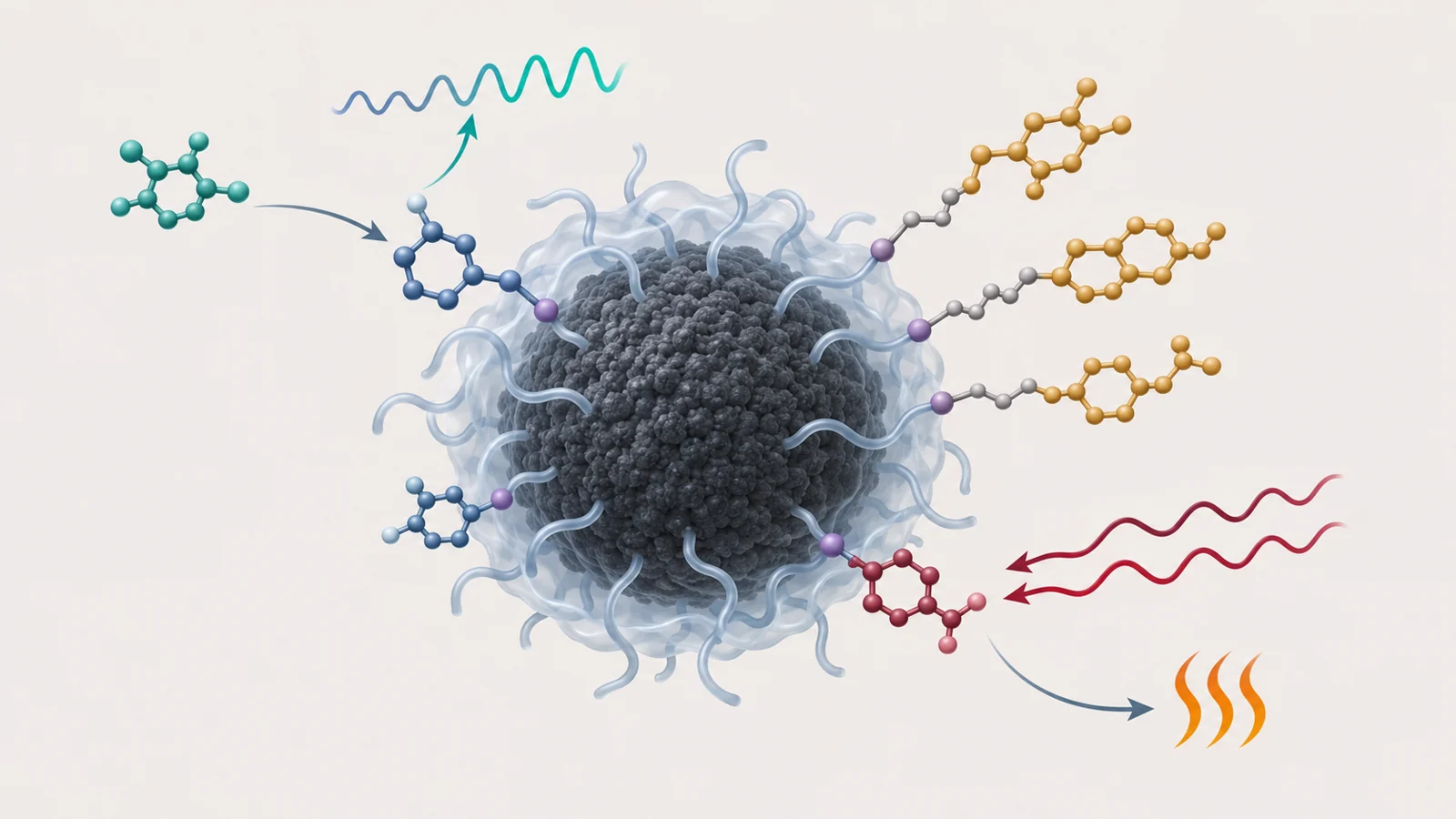
Carbon dots are tiny carbon-based particles that can emit light. A 2022 review describes how researchers adapt them for sensing, imaging, drug delivery and light-based treatments. Their flexibility is the appeal; it is also why a result from one recipe cannot stand in for every carbon dot.
A carbon core with a changeable exterior
The review describes carbon dots as structures with a carbon-rich core and polymer-like material at the surface, often without a sharp boundary between the two. The core can contain small crystalline carbon regions alongside less ordered material. Surface chains and chemical groups help determine how the particles interact with their surroundings.
Researchers can introduce groups such as hydroxyl, carboxyl, aldehyde, amino and sulfur-containing groups during preparation, or alter the surface afterward. Oxygen-containing groups tend to give dots neutral or negative charge, while nitrogen-containing groups can contribute positive charge. The mix also affects light emission, water solubility and the ways a dot can bind to another molecule.
This gives researchers several design choices, but “carbon dots” is not a single, uniform material. Different starting ingredients and preparation methods can produce particles with different structures and surfaces. Some methods use templates to help control size and shape; later modifications can add or change functions. The resulting formulation matters when interpreting both a useful effect and a safety finding.
A sensor reads a change in light
In the sensing examples, a carbon dot acts as a light-producing reporter. A target molecule interacts with the dot’s surface or an attached recognition group, and that interaction changes its optical signal. A detector can then measure the change.
One common design is “on-off” sensing: the dot fluoresces, then its emission dims when a target binds or otherwise interacts with it. The review describes metal ions such as iron and copper binding to surface groups, with electron transfer linked to reduced fluorescence. It also discusses glucose detection using dots prepared with boronic-acid groups, which interact with glucose and can change the particles’ emission.
Another design compares two emission bands instead of watching one signal fade. In a reviewed example, sulfur-doped dots and copper nanoclusters produced a two-color, or ratiometric, response to the insecticide dinotefuran. Comparing the two colors provides an internal reference, so the measured ratio changes with the target. These are sensing mechanisms described in the review; they do not, by themselves, establish a validated clinical test.
Imaging can use light or sound
For fluorescent imaging, the dots emit light that can help reveal where they collect in a sample. The review highlights interest in red and near-infrared emission because those wavelengths can travel farther through tissue and encounter less background fluorescence than shorter-wavelength light. Reported experimental examples include red- or near-infrared-emitting dots used to label cells and for imaging in living animals.
A different approach is photoacoustic imaging. Here, a particle absorbs light and converts some of that energy into heat, producing sound waves that can be detected. The signal is acoustic rather than the fluorescent glow itself. The review describes carbon-dot materials designed to absorb red or near-infrared light for this kind of imaging, including one example that was also studied for photothermal treatment. That pairing is an experimental demonstration, not a clinically validated imaging-and-treatment system.
Carrying a drug takes more than attaching it
A carbon dot can also serve as a carrier. Researchers have attached or loaded drugs, including anticancer medicines, to dots through binding or other forms of association. Some reviewed systems use chemical links that can be cleaved under particular conditions, with the aim of releasing a drug at a target site or in response to a stimulus.
Loading a medicine and controlling when it leaves are separate engineering problems. The review describes approaches that add polymers, hydrogels or porous materials to regulate release. In a porous material, a drug can move through a network of small channels; in a reservoir design, a surrounding membrane controls how quickly it diffuses out; in a swelling hydrogel, the polymer network’s mesh affects release. A carrier’s performance therefore depends on the whole delivery system, not simply on whether a drug can be attached to a dot.
Light-based treatments use different effects
In photothermal approaches, absorbed light becomes heat. In photodynamic approaches, light activates a process that produces reactive oxygen species—chemically reactive molecules that can damage nearby cells. They are distinct effects, even though both use light and may be combined in a single platform.
The review discusses carbon dots used as photothermal agents and as partners for photosensitizers, which are molecules that produce reactive oxygen under light. In one example, dots were linked to the photosensitizer Ce6; the review describes faster reactive-oxygen generation than with free Ce6 and greater light-triggered effects in the reported experiments. Other examples combine heat-producing dots with a photosensitizer so one light source can activate both processes. These are proof-of-concept strategies in the reviewed research, not evidence of established cancer treatment in people.
Safety depends on the formulation and exposure
The bacterial and animal findings illustrate why surface charge deserves attention, but they should not be collapsed into a single result. In the antibacterial examples, positively charged dots damaged the membrane of E. coli. The review also describes directly synthesized, strongly positive dots with a reported surface potential of about +45 millivolts. In rabbits, local eye administration of those dots was associated with opening of tight junctions in corneal epithelial cells and an antibacterial effect against Staphylococcus aureus. These were particular dots and a particular route of exposure.
A separate study examined five kinds of positively charged dots with similar measured surface potential but different charge density. Only the two with the highest charge density produced significant oxidative stress, release of the inflammatory marker IL-8 and mitochondrial dysfunction in the reported work; only those two induced airway inflammation in mice. One also increased allergen-related immune responses, airway inflammation and mucus production. The result shows why a broad label like “positively charged” may not capture the details that matter for safety.
Preparation and exposure conditions can also change what a safety test means. The review notes that concentration and illumination matter, and describes experiments in which fluorescent-lamp irradiation decomposed commercial dots and dots made from polyethylene glycol and glucose. In that work, cytotoxicity was mainly associated with small photolysis products released by the irradiated dots. A particle tested before and after illumination may therefore present different material to cells.
Safety assessments in the review include cell-viability tests and animal studies that examine blood measures, inflammation and organs. These kinds of results help characterize specific formulations under specific test conditions. They do not establish a universal toxicity profile for all carbon dots, nor do low-toxicity findings for one formulation settle the question for another.
How this could shape the future
If researchers can produce carbon dots with reliable, well-characterized surfaces and show that their behavior stays predictable under relevant exposure conditions, one material platform could support tools that report on molecules, help visualize biological processes or deliver a treatment while also showing where it goes. In practice, that could make experimental sensing and treatment systems easier to combine and study. The route from a promising particle to a dependable biomedical use depends on controlling what the particle is, what it carries and what happens to it in living systems. The review describes a range of possible functions; it does not establish a clinically validated all-in-one platform or human benefit.
Key Sources
- Primary source: Carbon Dots in Bioimaging, Biosensing and Therapeutics: A Comprehensive Review.
DOI or Journal Page · Review or Meta-Analysis · 2022-05-08
Keep exploring