
Research Signal – Source date 2022-05-01
Sleep can become longer even as the brain loses cells that help keep it awake. A study of Alzheimer disease and progressive supranuclear palsy found that patients with Alzheimer disease generally slept longer and maintained sleep better than patients with progressive supranuclear palsy in the clinicopathological comparison, despite showing greater loss across three wake-promoting brain regions.
That result is counterintuitive, but it is not evidence that neuronal loss improves sleep. Longer sleep is not automatically more restorative, and the comparison does not establish milder disease, better cognition, or a healthier brain. It points instead to a more specific possibility: different neurodegenerative diseases may disturb different parts of the machinery that balances waking and sleeping.
The comparison began with a sleep pattern
In the clinical comparison, patients with progressive supranuclear palsy slept for about 285 minutes on average, compared with about 400 minutes for patients with Alzheimer disease. Sleep maintenance was also lower in the progressive supranuclear palsy group, at about 61% versus 80% in the Alzheimer disease group. The progressive supranuclear palsy group spent more time awake after initially falling asleep and less time in NREM2 sleep.
The figures describe these study groups, not every person with either diagnosis. They also do not answer whether the additional sleep in the Alzheimer disease group was deep, refreshing, or helpful for daytime function. They describe sleep architecture and continuity, not a general measure of wellbeing.
The tissue findings added the puzzle. Across the three regions examined, the Alzheimer disease group had greater loss of wake-promoting neurons than the progressive supranuclear palsy group. A straightforward reading might expect more loss of wake-supporting cells to produce less wakefulness and therefore more sleep. The paper examined that possibility through measurements made during life and after death.
Sleep was recorded years before the brain tissue was counted
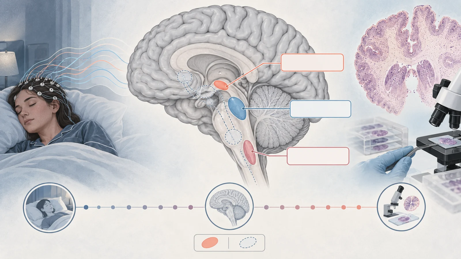
The larger study included 33 participants with Alzheimer disease, 20 with progressive supranuclear palsy, and 32 healthy controls. Participants were recruited through the University of California, San Francisco Memory and Aging Center between 2008 and 2020. Their sleep was assessed with electroencephalography or polysomnography, methods that record brain and body signals during sleep rather than relying only on a person’s estimate of how well they slept.
The clinicopathological analysis was narrower. It included 19 patients whose sleep measurements could be linked to postmortem examination: 10 with Alzheimer disease and 9 with progressive supranuclear palsy. The average interval between the sleep assessment and death was about 3.8 years. The disease groups also used different sleep-recording approaches, and participants continued their usual medication regimens during the sleep studies.
After death, researchers used immunohistological and stereological methods to examine three wake-promoting regions: the noradrenergic locus coeruleus in the brainstem, the orexinergic lateral hypothalamic area, and the histaminergic tuberomammillary nucleus. This chronology matters. The researchers were relating a sleep pattern observed at one point in a person’s life to neuronal counts obtained years later, not measuring sleep and cell number simultaneously.
Why losing wake-promoting cells could shift sleep
The three regions form an interconnected wake-promoting hub. In simple terms, they help provide the chemical signals that support alertness and keep the brain from remaining in sleep. If some of those cells are lost, the pressure favoring wakefulness may weaken. A weaker wake signal could allow longer sleep or make it easier to remain asleep, even though the underlying disease is progressing.
The observed correlations fit part of that interpretation. Greater numbers of lateral hypothalamic area and tuberomammillary nucleus neurons were associated with less total sleep, poorer sleep maintenance, more wake after sleep onset, and less NREM2 sleep. The relationships were substantial in this small group, with correlation values ranging from about −0.62 to −0.85 for several sleep measures. Locus coeruleus neuron counts were mainly associated with total sleep time and the delay before REM sleep.
But the balance is more complicated than a single wake switch. The study did not quantify the complementary sleep-promoting neurons or the circadian-regulating system. Fewer wake-promoting cells could be one part of the explanation, while changes elsewhere in the sleep-wake network could shape the final pattern. The authors specifically raised the possibility that sleep-promoting neurons might be selectively vulnerable in progressive supranuclear palsy, but that possibility remained untested in this analysis.
Tissue patterns crossed diagnostic boundaries
The researchers also combined the neuronal and pathological measurements from all three regions and used k-means clustering. This produced two groups called Alzheimer-predominant and progressive-supranuclear-palsy-predominant. The groups contained 11 and 8 participants, respectively, and were defined by their tissue measurements rather than by simply sorting everyone according to the clinical diagnosis.
The Alzheimer-predominant group had higher total sleep time, better sleep maintenance, less wake after sleep onset, and more NREM2 sleep than the progressive-supranuclear-palsy-predominant group. The clustering therefore reproduced the broad sleep contrast while showing that the biological pattern was not perfectly identical to the disease label. The paper notes that some pathological profiles crossed those diagnostic boundaries, including PSP pathology that clustered with the Alzheimer-predominant group.
This matters because a diagnosis names a broad disease pattern, while sleep symptoms may depend on which connected circuits have been affected in an individual. The result is not a validated classifier and does not show that tissue measurements can identify a person’s disease or predict their future sleep. It does suggest that mixed or uneven pathology may help explain why people carrying the same diagnosis can have different sleep problems.
What the design can and cannot connect
The most important constraint is the size of the matched sample: 19 patients. A small group can reveal a strong association while still giving an unstable estimate of how widely the pattern applies. The participants were also drawn from one specialist center, and all patients in the clinicopathological analysis were White, limiting how confidently the findings can be extended to other populations.
The timing creates another boundary. A neuronal count taken years after a sleep recording cannot show exactly what the brain looked like on the night of that recording. The researchers adjusted for the interval between assessment and death, but adjustment does not turn separate measurements into a simultaneous experiment. Different recording methods across the disease cohorts also make direct comparisons less clean than they would be under one shared protocol.
Medication is part of the context as well. Because participants continued their usual regimens, the results describe sleep in the conditions in which these patients were living, not the isolated effect of neuronal loss. The study was observational, so it cannot establish that losing a particular group of neurons caused an individual’s sleep pattern.
Finally, the missing sleep-promoting and circadian measurements leave the proposed circuit balance incomplete. The data support a relationship between wake-promoting neuronal counts and sleep phenotypes, but they do not identify the whole causal pathway. Longer sleep in the Alzheimer disease group therefore should not be read as evidence of improved sleep quality, less severe neurodegeneration, or a treatment effect.
What this could change
The practical promise is more appropriately matched help for people whose neurodegenerative disease disrupts wakefulness or sleep in different ways. If future research confirms that Alzheimer disease and progressive supranuclear palsy damage distinct parts of the sleep-wake network, care could eventually be designed around the affected circuitry rather than treating every sleep complaint as the same problem.
Reaching that point would require confirmation of the circuit differences in larger and more diverse groups, along with evidence that acting on those differences safely improves patients’ symptoms. The 2022 study supplies a clinicopathological clue toward that goal, not a drug recommendation or a proven treatment benefit.
Key Sources
- Primary source: Subcortical Neuronal Correlates of Sleep in Neurodegenerative Diseases.
DOI or Journal Page · Peer-Reviewed Paper · 2022-05-01
Keep exploring