
Research Signal – Source date 2026-05-01
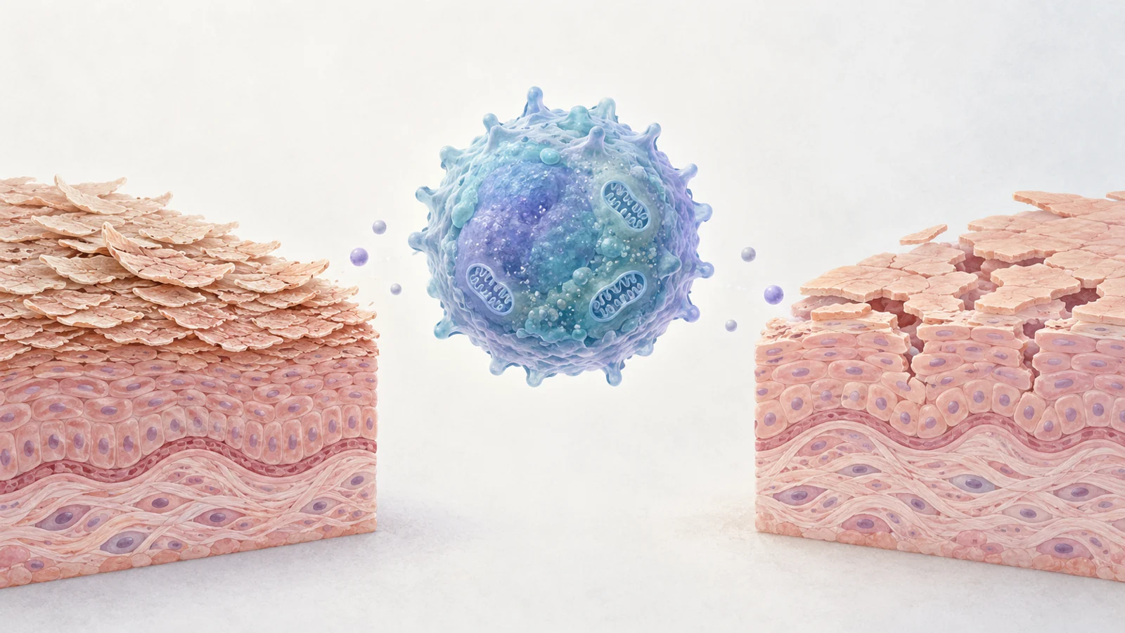
Aging immune cells are often described as less capable. Yet some T cells with features of aging can remain metabolically active and release inflammatory substances. In skin, that combination may help explain how inflammation persists even as immune function changes.
A review published May 1, 2026, examines this possibility in psoriasis, atopic dermatitis, rosacea, and seborrheic dermatitis. Its authors report searching peer-reviewed English-language papers through February 2026. They bring together observations in human skin, studies of treatments that block inflammatory pathways, small clinical reports, and experiments that target senescent cells more directly. The evidence points to a useful question: can treatment quiet the substances these cells release, or reach the aging cells themselves?
When an aging T cell stays inflammatory
T cells help coordinate immune responses. With age or ongoing stress, some acquire features associated with immunosenescence, a gradual change in their function. The review describes a familiar marker pattern: reduced CD27 and CD28, alongside increased CD57 and KLRG-1. Telomere shortening, mitochondrial dysfunction, and changes in cell-cycle control can also be part of the picture.
The apparent paradox is that reduced immune competence does not necessarily mean silence. A senescent T cell may stop dividing while continuing to release inflammatory signals. This collection of secreted factors is called the senescence-associated secretory phenotype, or SASP. Depending on the cell and tissue, it can include cytokines, chemokines, and molecules involved in tissue remodeling. Those signals may recruit or activate other immune cells and keep nearby tissue inflamed.
A marker alone cannot tell the whole story. CD57 or KLRG-1 may help identify a cell with senescence-associated features, but the review’s account also depends on function: whether the cell has stopped proliferating, what it secretes, and how it interacts with surrounding cells. Senescence can also be confused with exhaustion. Exhausted T cells are commonly described by inhibitory receptors such as Tim-3 and TIGIT and impaired effector activity. Senescent T cells may share some features with terminally differentiated T cells, but the review emphasizes their potential to remain an active source of inflammatory signals.
Psoriasis links stressed T cells to plaque inflammation
In psoriasis, the review connects senescence-associated T-cell findings in lesions with the familiar cycle of thickened, scaly plaques. It cites human observations of increased CD4 T-cell subsets expressing the cell-cycle inhibitors p16 and p21 in lesional skin. Other reported observations describe more terminally differentiated or senescence-associated T-cell populations in people with psoriasis, particularly with longer disease duration. These findings show that such cells are present; by themselves, they do not establish that the cells initiate the disease.
The proposed mechanism gives them a way to contribute. In the review’s account, IL-23 can activate JAK and STAT3 signaling in T cells, supporting production of IL-17A and IL-22. IL-17A can amplify inflammation, while IL-22 can act on keratinocytes, the main cells of the outer skin layer, and promote abnormal growth and differentiation associated with thickened plaques and scale.
Other secreted signals may help recruit more inflammatory cells. The review describes NF-κB activity and the chemokine CCL20 as part of a route that attracts CCR6-bearing Th17 cells and dendritic cells into skin. More arriving immune cells can add to local inflammation, giving the tissue a way to sustain the response. These pathways connect senescent T cells to plaque inflammation in the review’s proposed model, alongside other immune and skin-cell processes involved in psoriasis.
In eczema, inflammation can weaken the barrier and intensify itch
Atopic dermatitis, often called eczema, follows a different pattern in the review. Type 2 inflammatory signals such as IL-4 and IL-13 are linked to reduced production of important skin-barrier proteins, including filaggrin, loricrin, and involucrin. A weakened barrier can leave skin dry and more vulnerable to irritation.
The review describes a feedback loop between immune signals and the skin. T-cell-derived IL-4 and IL-13 can affect keratinocytes and impair barrier maintenance. Damaged keratinocytes, in turn, release signals that may stimulate further type 2 activity. IL-31 is associated with itch and is also discussed as part of an inflammatory loop. Together, barrier disruption and persistent itch help explain how the proposed T-cell contribution could matter to daily symptoms, rather than simply adding another immune pathway to a list.
The review also discusses IL-31 as a possible contributor to senescence-associated changes in T cells. That link is part of the proposed biology, not proof that senescent T cells account for eczema in general. Atopic dermatitis involves more than one process, and the review does not position T-cell aging as its sole cause.
Rosacea and seborrheic dermatitis have distinct proposed routes
The proposed connections are less developed for rosacea and seborrheic dermatitis. In rosacea, recurrent flushing and persistent facial redness are associated with neurovascular changes and innate immune activity. The review notes a proposed role for a SIRT7–TLR2–NF-κB pathway in older people, while also describing the relationship between aging and rosacea as poorly understood.
Seborrheic dermatitis is discussed through interactions between skin cells, Malassezia, a skin-associated fungus, and immune signaling. The review proposes that keratinocytes respond to Malassezia by releasing IL-23, while T cells respond to fungal metabolites. In that account, NF-κB and STAT3-related signaling can support inflammatory substances including IL-17A, IL-1β, and TNF-α. The resulting inflammation is linked to redness and papules, while IL-17A is proposed to affect keratinocyte differentiation and contribute to greasy scale.
These accounts are not equally established. The review describes rosacea and seborrheic dermatitis treatment evidence largely as case reports or small case series. Such observations can suggest a possibility, but they do not show that the same mechanism operates across patients or that a treatment effect will be consistent.
Existing drugs interrupt signals; direct cell targeting remains experimental
Some treatments already in use act on inflammatory signals discussed in the review. For atopic dermatitis, dupilumab blocks IL-4 receptor alpha, a shared component of the receptors for IL-4 and IL-13. For psoriasis, drugs that block IL-17A act on a downstream inflammatory signal. The review summarizes clinical evidence for these approaches and discusses other drugs that inhibit signaling pathways such as JAK–STAT.
These treatments can reduce disease activity by interrupting signals, but their effects do not demonstrate that T-cell aging has been reversed. Blocking a cytokine or signaling hub can change what happens in the tissue without removing or reprogramming the cell that produced the signal. The review frames this as an open therapeutic distinction: intercept inflammatory outputs, or target senescent cells and the processes that sustain them.
More direct approaches remain at an earlier stage in the evidence described. In a psoriasis-like mouse model, a topical BCL-2 inhibitor was reported to reduce senescent-cell proportions and SASP-related signals in lesions. Other experiments in cells or mice associated caffeine exposure or time-restricted feeding with fewer senescence-associated T cells and reduced psoriasis-like inflammation. These findings are reasons to investigate direct targeting; they do not establish durable remission or safety for such treatment in people.
What the evidence can tell us
The review combines evidence that answers different questions. Human lesion observations show that T cells with senescence-associated features can be found in psoriatic skin. They do not, on their own, determine whether those cells cause disease, follow chronic inflammation, or do both. Clinical studies of drugs that block inflammatory targets can show whether those treatments help with disease outcomes; they do not establish that the drugs reverse T-cell senescence.
Small reports in rosacea and seborrheic dermatitis offer early clinical observations, not a shared treatment rule. Cell and animal experiments can test whether more direct interventions change senescent-cell features or disease-like inflammation in controlled settings. They cannot establish what those interventions would do over time in people. The review’s search period also matters: it covers literature through February 2026, while the article itself was published on May 1, 2026.
Could quieting aging T cells extend remission?
If researchers can identify which senescent T cells actively sustain skin inflammation and safely interrupt that activity, treatment might eventually reach a source of recurrence as well as its inflammatory outputs. That could add a route toward longer-lasting control for people whose disease remains active despite blocking individual signals. The possibility depends on establishing that the cells are causal in each disease, that they can be targeted without harmful effects, and that results from preclinical models translate to people. For now, the review presents that goal as a research direction, not a demonstrated clinical outcome.
Key Sources
- Primary source: T Cell Immunosenescence in Inflammatory Skin Diseases: Pathogenesis and Therapeutic Targets.
DOI or Journal Page · Review or Meta-Analysis · 2026-05-01
Keep exploring