
Research Signal – Source date 2026-06-17
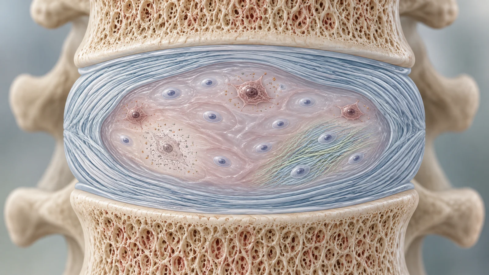
Removing dysfunctional, aging cells from a damaged spinal disc might reduce one source of ongoing harm. By itself, though, clearance would not replace cushioning material that has already broken down or restore the remaining cells’ ability to make healthy tissue. That is the logic behind a proposed regenerative sequence in a review published June 17, 2026: clear harmful cells, prime the cells that remain, then seed the tissue with replacement cells if needed. The sequence is a research proposal, not a demonstrated treatment for people.
Intervertebral discs sit between the bones of the spine and help cushion them. Degeneration can contribute to low back pain, but current care described in the review mainly relieves symptoms or restores mechanical stability. The authors argue that biology deserves attention alongside mechanics because changes in disc cells may help drive tissue damage forward.
Senescent cells can change their neighborhood
A senescent cell has entered a persistent altered state. Rather than dying or functioning normally, it stops dividing and loses some of its capacity to maintain healthy tissue. In a disc, the review describes these cells as producing less of the extracellular matrix, the material that gives the tissue much of its structure and cushioning, while releasing a mixture of signaling molecules and enzymes.
That mixture is called the senescence-associated secretory phenotype, or SASP. It includes inflammatory signals and matrix-degrading enzymes. The review describes how these substances can weaken the surrounding matrix and alter the local tissue environment. Some signals may also push nearby disc cells toward senescence, extending the problem beyond the cells that first changed.
The authors discuss the CXCL10/CXCR3 signaling pathway as one proposed route for this spread. In the cited mechanistic work, CXCL10 from senescent cells activates p38 MAPK in neighboring cells and raises p16, a marker associated with senescence. Other signals, including CCL2 and IL-8, have also been implicated in this process. These findings help explain how cell behavior could reinforce tissue deterioration; they do not show that every senescent cell acts identically or that this pathway alone accounts for disc degeneration.
The resulting feedback may matter in a tissue that already has limited capacity to recover. In the review’s account, inflammatory signals and proteases can compromise matrix integrity and affect the disc’s mechanical properties. Senescence-associated metabolic changes may add further stress to the local environment. As the tissue environment worsens, its cells may find it harder to sustain the matrix that normally supports the disc.
An aging signal is not yet a reset button
Epigenetic marks help regulate which genes are active without changing the DNA sequence itself. The review describes DNA methylation, chemical marks on DNA, alongside histone modifications, marks on the proteins around which DNA is organized, and non-coding RNAs that influence gene activity. Together, changes in these systems may both reflect aging and contribute to the loss of a healthy nucleus pulposus cell identity. The nucleus pulposus is the soft central region of the disc.
One human-tissue finding summarized by the authors gives the “epigenetic clock” idea a concrete basis. Severely degenerated discs had a higher measured epigenetic age than the donors’ chronological age, and the reported age acceleration was greater in samples with more advanced degeneration. This is an association in human samples. It does not establish that clock changes start the disease, or that changing a clock reading would restore disc function.
The gene-level findings help show why the clock concept could matter biologically. The review reports that promoter regions for ACAN, which encodes the matrix protein aggrecan, and SOX9, a regulator of disc-cell identity, can carry unusually high DNA methylation in degenerated tissue. Such marks may impede gene activity and reduce matrix production. It also describes higher EZH2 expression and enrichment of the repressive histone mark H3K27me3 at promoters of genes including FOXF1 and KRT19, which are associated with nucleus pulposus identity. These observations connect epigenetic regulation to cell function, but evidence that the marks can be changed is not the same as proof that the whole degenerative process can be reversed.
Causation remains a central open question. The review notes that direct longitudinal and intervention studies tracking disc epigenetic clocks are sparse. For example, work summarized by the authors found benefits from the DNA methylation inhibitor 5-azacytidine in an experimental cell setting, but did not directly show restoration of SOX9 or other master identity genes. A separate mouse study of senolytic treatment reduced senescent-cell burden and structural degeneration, while whether disc clock marks changed was not examined. The clock is therefore both a possible measurement tool and a possible therapeutic target, but those roles still need to be disentangled.
Three strategies with different jobs
The review groups emerging approaches around three complementary aims. Senolytics seek to remove senescent cells. Senomorphics aim to suppress harmful secretions without killing the cells. Either approach could reduce a source of damaging signals, but clearance or suppression alone does not replenish degraded matrix or teach a dedifferentiated cell to make it again. This is why the authors look beyond cell removal as a standalone strategy.
“Prime” means trying to restore a healthier gene-activity pattern in cells that remain. The review discusses small-molecule epigenetic remodeling, including EZH2 inhibitors that reduce the repressive H3K27me3 mark. In rat models, local use of such inhibitors was reported to attenuate degeneration and reactivate FOXF1 and KRT19 expression. More precise CRISPR-dCas9 tools could, in principle, add or remove epigenetic marks at selected genes without changing the DNA sequence. The review describes this as an emerging approach, with limited disc-specific evidence and practical challenges that include delivery, unintended binding and immune response.
“Seed” means supplying cells or cell-derived material that could help restore tissue-building capacity. The review discusses induced pluripotent stem cell-derived notochordal cells and exosomes from induced pluripotent stem cell-derived mesenchymal cells. It summarizes a porcine model in which notochordal cells reduced degeneration. Such work is preclinical: it does not establish that transplanted cells will retain a useful identity in a human disc or rebuild its matrix safely.
The authors’ “clear, prime, then seed” sequence puts the approaches in a proposed order. First reduce harmful senescent-cell activity, then encourage surviving cells toward a healthier identity, and finally add replacement cells where needed. The rationale is that replacement cells may face a damaged environment unless harmful signals are addressed, while cell clearance alone leaves lost tissue-building capacity unresolved. The combined sequence has not been shown to regenerate a human disc or relieve patients’ pain.
The route from a model disc to a human treatment
Much of the evidence reviewed comes from cell experiments and animal models. Rodent discs differ from human discs in anatomy, cellular composition, mechanical loading and patterns of degeneration. One difference the authors emphasize is that rodents retain notochordal cells throughout life, while humans largely lose them after adolescence. A promising result in a rodent therefore may not translate to a human disc, and the review calls for more human-relevant models and testing in larger animals.
Delivery and safety add further hurdles. Disc tissue is difficult to reach, and a useful treatment would need to act in the right cells and tissue region. Broad epigenetic changes could affect more than the intended disc-cell genes; targeted editing faces questions about delivery, off-target binding and immune response. Cell therapies also need to address the possibility of residual undifferentiated cells, unstable cell identity in the disc environment and demanding manufacturing. These are design and translation problems, not details that a promising cell or animal result can settle by itself.
Clinical evidence must be kept in its proper lane. The review notes that some candidate drugs have been used or studied for other conditions, but this does not show that they repair discs. It reports no dedicated clinical trials for IDD among the senolytic approaches it summarizes, and no disc trials for the CRISPR-dCas9 strategy. Early human studies, if warranted after further work, would need to establish safety and show that the intended biological target was engaged. They would also need to assess whether disc tissue is restored and whether patients experience meaningful improvement in pain and function.
What disc regeneration could mean for low back pain
A treatment that safely restores disc-cell function and the cushioning matrix could give some patients a way to address degeneration itself, beyond symptom relief or mechanical stabilization. That possibility depends on demonstrating real tissue restoration, meaningful patient benefit and acceptable safety in human studies. The proposed sequence offers a way to organize that research; it is not yet evidence that a damaged spinal disc can be biologically reset.
Key Sources
- Primary source: Resetting the epigenetic clock: cellular senescence and regenerative strategies in intervertebral disc degeneration.
DOI or Journal Page · Review or Meta-Analysis · 2026-06-17
Keep exploring