
Research Signal – Source date 2026-04-01
In this mouse study, an anti-inflammatory compound increased sleep during the animals’ usual rest period, even though measured amyloid plaques did not shrink. The result raises a focused question: can sleep disruption improve while Alzheimer’s-related brain pathology remains? The experiment offers evidence that it can in these mice, while leaving open what caused the change and whether it could matter for people.
The paper, published April 1, 2026, studied knock-in mice carrying three human mutations in the amyloid precursor protein gene. These mutations model inherited forms of Alzheimer’s-related amyloid pathology. Unlike models that overproduce the protein, this model retains its normal gene location and expression pattern. The researchers followed sleep and behavior in separate groups of mice, then tested a drug in a separate group of older animals. Those parts of the study answer related but different questions: how sleep changed with age, and whether treatment altered sleep at that later age.
Female mice began sleeping less during their rest period
Mice are generally more active during the dark phase and rest during the light phase. Researchers used pressure-sensing floors beneath the home cages to estimate sleep: sustained stillness, with breathing-related movement, counted as sleep; active movement counted as wake. The system allowed repeated monitoring in familiar cages, but it did not measure human-style sleep stages.
In the longitudinal group, sleep was measured at ages from 2 to 19 months. Female mice with the mutations began sleeping less during the light phase at about 10 months compared with female controls. The gap persisted at later measurements; by 19 months, mutant females slept 8.8% less during that phase. The highlighted genotype difference was not detected in males, whose light-phase sleep remained similar to that of male controls across the measured ages.
The rhythm findings had their own timing. In females, activity rhythm amplitude, a measure of the difference between activity peaks and troughs, was lower in mutant mice at 8 and 10 months. Fragmentation increased later: mutant females showed more frequent transitions between rest and activity at 15, 17, and 19 months. Their day-to-day rhythm stability was also lower at several ages. These measures describe different features of the rest-activity pattern; they do not amount to one single measure of sleep quality.
The behavioral results were also specific. Mutant mice learned a water-maze task and retained the learned location similarly to controls in the reported comparisons. Older mutant females made more errors when the task rules were reversed, a test of adapting to a changed goal. That selective difference is not evidence of a general learning or memory failure. Nest-building performance declined with age in both sexes, with deficits emerging in mutant mice at later ages. Neither the maze nor nesting results show what the drug changed, because the behavioral and treatment questions came from separate cohorts.
A week-five difference emerged during treatment
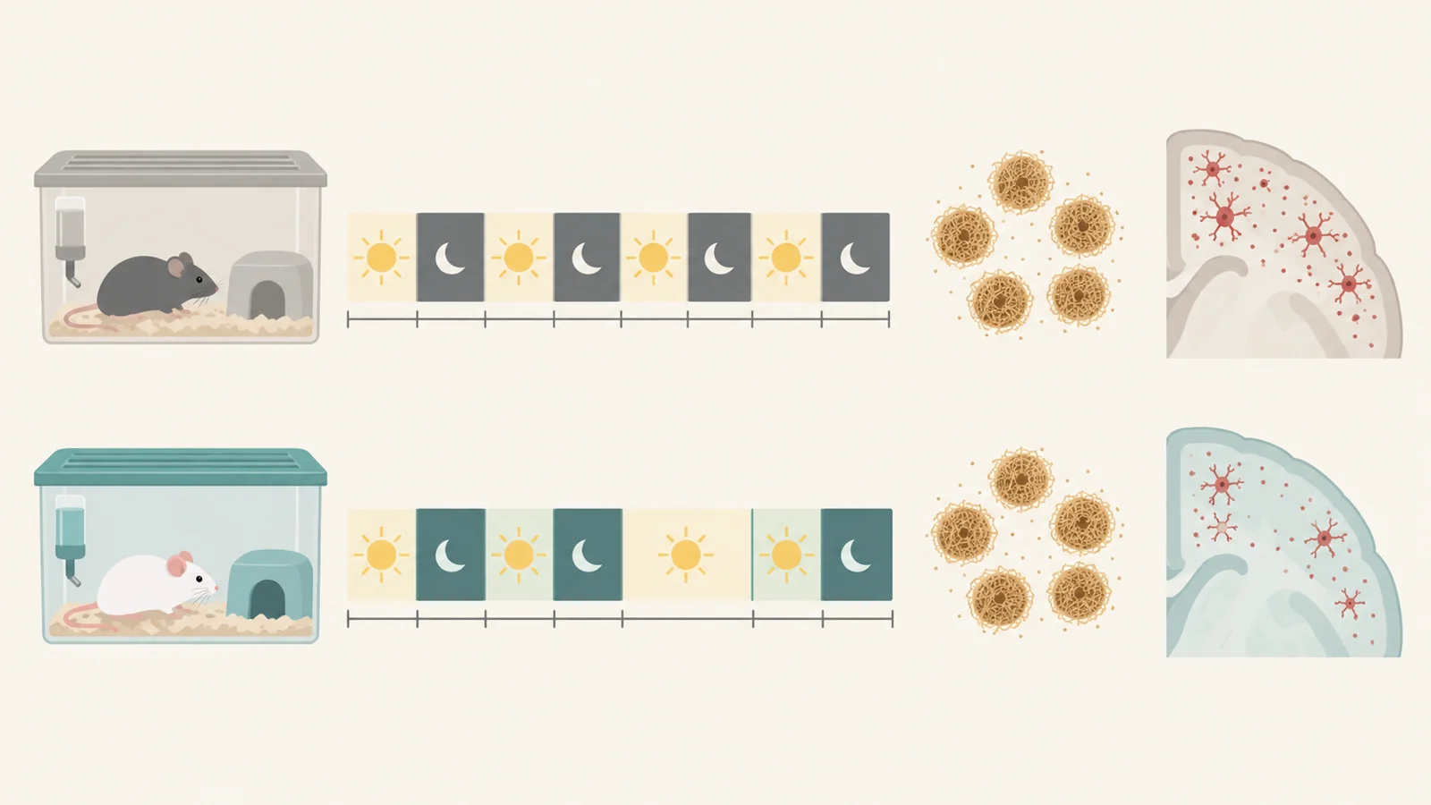
The intervention group consisted of older mutant mice, about 15 months old at the start. After baseline sleep recordings, mice were assigned to receive MW151 or saline vehicle by mouth every other day for six weeks. Sleep was recorded before treatment and again during weeks three and five. Researchers prespecified the week-five comparison, when both groups had undergone the same handling and dosing schedule.
At baseline, the assigned groups had similar sleep and rhythm measures. There was little separation at week three. By week five, mice receiving MW151 spent more time asleep during the light phase than vehicle-treated mice. The treatment effect was statistically significant and had a large reported effect size. The average level of activity across the daily rhythm also fell, consistent with more rest during the light phase, while rhythm amplitude did not change.
The result was limited to particular outcomes. Dark-phase sleep, rhythm fragmentation, activity rhythm amplitude, and total 24-hour sleep did not differ significantly between treatment groups in the reported comparison. Total daily sleep showed a medium-sized estimated effect, but it did not meet the study’s statistical threshold. The light-phase sleep response did not differ statistically by sex within the mutant group. That does not establish that every treatment effect was identical in females and males; it means the analysis did not detect a treatment-by-sex difference.
Sleep changed alongside one inflammatory marker
At week six, the researchers measured inflammatory proteins in the brain’s outer tissue, the cortex, and assessed amyloid in tissue samples and by plaque staining. MW151 reduced cortical TNF-α, an inflammatory signaling protein. The study did not detect a treatment effect on IL-1β or the other measured cytokines and chemokines. Nor did it detect a change in measured amyloid fractions or plaque counts in the cortex and hippocampus.
The sleep and TNF-α findings are consistent with the drug engaging an inflammatory target while sleep changed, without reducing the measured amyloid burden. They do not show that TNF-α alone caused the sleep response. The study did not identify the brain circuit connecting cortical inflammation to sleep regulation, and other biological changes could have contributed. Earlier differences in sleep among females also cannot be treated as predictors of drug response: the age-tracking and treatment experiments used separate cohorts.
The sex pattern deserves similar care. Sleep disruption was more evident in mutant females, but researchers did not directly measure reproductive hormones or cycling. The timing overlaps with reproductive aging in mice, which makes hormonal influence a possibility, not a finding of this experiment. Other sex-related differences could also be involved.
Could Alzheimer’s-related sleep loss be treated separately from plaques?
If controlled studies eventually show a similar effect in people, a treatment aimed at inflammatory signaling could increase nighttime sleep even if amyloid plaques remain. That would be a practical benefit for people with Alzheimer’s-related sleep disruption. This study establishes only more light-phase sleep in mice at week five, without a significant increase in total daily sleep or evidence of broad rhythm restoration; it provides no evidence of benefit in people.
The paper also discloses that one author holds patents covering MW151 and helped found a company formed to commercialize it, and another co-founded the company that makes the sleep-monitoring technology used. These interests do not determine whether the results are valid, but they are relevant context for readers weighing a small-animal study and its proposed therapeutic implications.
Key Sources
- Primary source: Glial cytokine modulation improves sleep and circadian disruption in female SAA knock-in mice of Alzheimer's-related pathology.
DOI or Journal Page · Peer-Reviewed Paper · 2026-04-01
Keep exploring