
Research Signal – Source date 2026-04-01
Chronic kidney disease (CKD) often advances through fibrosis, the buildup of scar-like material that disrupts kidney tissue. It can involve glomerulosclerosis, tubular atrophy and scarring between the tubules. As functioning tissue is lost, kidney function can decline toward kidney failure, when some people need dialysis or a transplant.
A review published on April 1, 2026, examines a possible contributor to that progression: cellular senescence. Senescent cells have stopped dividing after stress or damage, yet some remain active and release signals that affect surrounding tissue. The central question is whether these cells help drive chronic kidney damage, or mainly accumulate because damage is already underway. The evidence points to a possible two-way relationship, but does not settle which role predominates in patients.
A short-lived response can become a lasting problem
Senescence is not always harmful. After an injury, a temporary senescence response can help contain damage, support wound healing and allow immune cells to clear affected cells. In the kidney, natural killer cells can recognize and eliminate senescent tubular cells through an immune process described in the review. Timely removal helps keep this response temporary.
The concern is persistence. When stress continues or the immune system cannot keep pace with the production of senescent cells, they may accumulate. These cells resist normal renewal and can release a collection of inflammatory and other signals known as the senescence-associated secretory phenotype, or SASP.
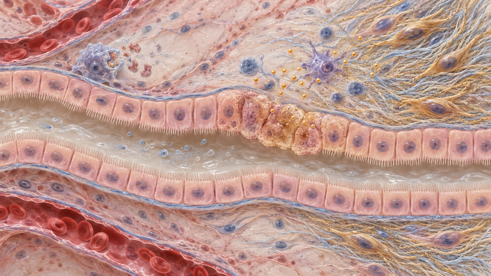
Those signals may affect nearby cells, and the review describes two routes for senescence to spread. In paracrine signaling, secreted factors act on cells nearby or travel through body fluids. In juxtacrine signaling, cells influence neighbors through direct contact. A response that began in a limited group of damaged cells could therefore alter the behavior of a wider area of tissue.
The triggers can vary by kidney cell type. Podocytes, for example, divide slowly, so stressors such as oxidative damage, low oxygen or uremic toxins may matter more to their senescence than the exhaustion of repeated cell division. Different triggers can also produce different senescent-cell features. That variation matters when researchers try to identify the cells or choose a treatment aimed at them.
How injured tubules may add to scarring
The review traces a route from persistent tubular injury to fibrosis. Proximal tubular epithelial cells that remain stressed may become stuck in a stage of the cell cycle called G2/M arrest. In that state, they can release signals through JNK-dependent pathways that promote fibrosis in the surrounding tissue.
Over time, this kind of maladaptive repair can interfere with the restoration of the tubule lining. Inflammation and signals from injured or senescent cells can encourage the accumulation of extracellular matrix proteins, including collagen and fibronectin. As that material builds up between tubules, tubulointerstitial fibrosis can impede kidney function. The review also discusses the transition from acute kidney injury to CKD as a setting where persistent cell-cycle arrest and faulty repair may contribute to lasting damage.
CKD may also create conditions that further stress kidney cells. The review describes protein-bound uremic toxins, including indoxyl sulfate and p-cresyl sulfate, as potential contributors to oxidative stress, DNA damage responses, mitochondrial dysfunction, cell-cycle arrest and SASP signaling. This gives the proposed relationship a feedback quality: injury may promote senescence, while persistent senescence may help sustain inflammation and fibrosis.
Human observations and animal interventions answer different questions
Human kidney studies summarized in the review have found senescence-associated markers, including p16 expression and SA-β-Gal activity, across CKD and several kidney conditions. Some studies also link higher marker levels with more fibrosis or disease progression. These observations make senescence relevant to investigate, but markers alone cannot show whether senescent cells caused the damage, developed in response to it, or both. No single marker reliably identifies every senescent cell.
Animal experiments offer a different kind of evidence. In models where senescent cells were genetically reduced or removed, kidney function improved and fibrosis fell. These interventions strengthen the case that senescent cells can contribute to kidney injury under experimental conditions. They do not determine how much they drive CKD in people or establish that a particular treatment will be safe and effective for patients.
Together, the human findings and animal experiments support a plausible role in disease progression while leaving the cause-and-consequence question open in patients. That distinction matters for treatment: a cell state can be associated with worsening disease and still require careful testing before targeting it improves a patient’s kidney outcomes.
Removing cells and quieting their signals carry different tradeoffs
Senolytics are designed to eliminate selected senescent cells. Senomorphics aim to suppress harmful features, such as SASP signaling, while leaving the cells in place. The first strategy targets the cells themselves; the second targets some of their effects.
These approaches bring different treatment burdens. A senolytic might be given intermittently, while a senomorphic that needs to keep suppressing cell behavior could require ongoing treatment. Clearing cells may also have lasting consequences that are difficult to predict, because senescence can serve useful roles in repair and other processes. Suppressing signals may be less drastic, but SASP factors also have physiological functions, including roles in immune surveillance.
The right approach may depend on which cells are affected and when treatment is given. The review notes that safety and effectiveness could differ across senescent-cell types and CKD stages. Advanced CKD can come with reduced regenerative capacity and greater frailty, while kidney impairment can change the handling of drugs cleared through the kidneys. Immune dysfunction and blood abnormalities may further complicate safety. The review notes that navitoclax can cause low neutrophil counts and bone effects, illustrating why experimental promise does not settle the risk-benefit balance for people with CKD.
A human treatment signal is still several steps from kidney benefit
The review describes a human study in diabetic kidney disease in which treatment with dasatinib and quercetin reduced circulating SASP factors and the abundance of senescent cells in adipose tissue and skin. This is a treatment-related change in measured markers outside the kidney. It is not evidence that senescent cells were cleared from kidney tissue, that kidney function was preserved, or that CKD progression slowed.
The review notes that clinical trials of senescence-targeting treatments remain limited relative to laboratory and animal research, with additional trials of dasatinib and quercetin and fisetin underway at the time it was published. For people with CKD, drug clearance, frailty and immune or blood-related vulnerabilities make kidney-specific safety part of the question, alongside whether a treatment changes disease outcomes.
To interpret a future trial, researchers would need measures that connect a treatment to meaningful kidney changes. The review discusses kidney fibrosis measures, circulating or urinary senescence-related factors, extracellular-vesicle signatures and standard measures such as estimated glomerular filtration rate (eGFR). Changes in circulating markers could help show that a treatment affects its intended biology; they would still need to be considered alongside kidney function and patient safety.
Finding the harmful cells remains a challenge
Senescence is a complex state, and markers such as p16, p21 and SA-β-Gal are not individually definitive. Inflammation markers are also insufficient on their own because many can appear for reasons other than senescence. A useful test would need to distinguish persistent, harmful senescence from temporary repair responses and other stressed or resting cell states.
The review describes research using artificial intelligence to analyze features such as nuclear shape, chromatin texture and loss of LMNB1, alongside molecular and clinical data. These approaches may help quantify senescence in tissue images and compare patterns across samples. Combining image-derived measures with circulating or urinary markers could eventually make assessment more practical, but the review presents these tools as developing research approaches rather than routine kidney tests. Urinary exosomes are a possible source of information; standardized collection and characterization remain necessary.
What slowing kidney scarring could mean for patients
If researchers can reliably identify persistent senescent cells that are contributing to kidney scarring, and trials show that targeting them is safe and improves kidney outcomes, senescence-directed treatment could give some people with CKD another way to slow loss of kidney function. That possibility depends on solving two linked problems: finding the right cells and showing that changing them benefits patients, rather than only shifting markers. Until then, senescence is a promising explanation and treatment target under study, not an established therapy for CKD.
Key Sources
- Primary source: Chronic Kidney Disease and Cellular Senescence.
DOI or Journal Page · Review or Meta-Analysis · 2026-04-01
Keep exploring