
Research Signal – Source date 2026-03-31
A cell can stop dividing and still influence the tissue around it. Stressed cells in this state, called senescent cells, release a changing mix of chemical signals. During fracture repair, some of those signals may help organize early healing; if senescence and its secretions persist, they may instead contribute to inflammation, scarring and delayed union.
A review published on March 31, 2026, brings this tension into focus by arranging senescence-associated secretions across the overlapping stages of fracture healing. Its central idea is that timing, location and the mix of signals matter. That idea helps explain why removing senescent cells might help in some experimental settings yet could disrupt repair if done at the wrong point.
Repair unfolds through overlapping stages
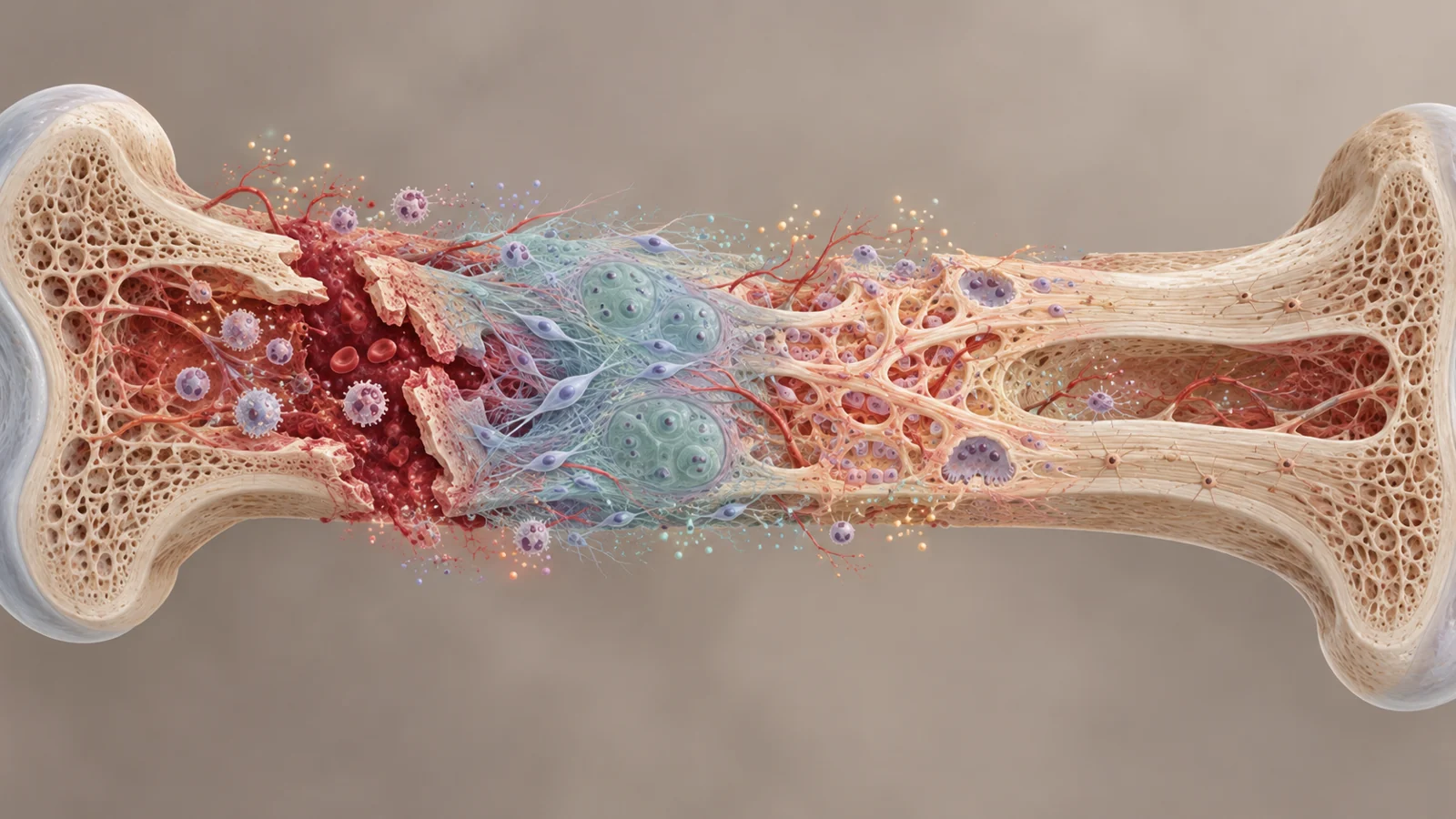
A broken bone first fills with a blood clot, or hematoma. The clot provides a temporary structure and a hub for cells and chemical messages. Inflammation brings immune cells that clear debris and help initiate repair. As that response settles, connective tissue and cartilage build a soft callus across the break. The callus later hardens as cartilage is replaced by bone, followed by remodeling that reshapes the new bone.
These stages overlap, and the local conditions influence how bone forms. With good blood supply and stability, bone-forming cells can deposit new bone directly along the periosteum, the tissue covering the bone. In less stable or poorly supplied regions, a cartilage bridge can form first and later be replaced by bone. Both routes can contribute to repair in the same fracture.
The review uses this sequence to organize the senescence-associated secretory phenotype, or SASP: the collection of substances senescent cells release. The SASP is not a fixed recipe. Its possible contribution changes as immune recruitment gives way to callus formation, cartilage replacement and structural remodeling.
The molecule does not name its maker
A key interpretive problem is that many SASP-associated molecules also appear in the ordinary response to injury. A fracture hematoma contains signals from immune cells, platelets, blood cells and activated stromal cells. Finding an inflammatory molecule there cannot, by itself, show that a senescent cell produced it.
The review notes that senescent cells may add a distinct layer to this broader injury response, but the source of shared molecules needs to be tied to evidence of senescence, such as cellular markers and cell-specific analyses. In mouse fracture studies, senescent cells have been observed among mesenchymal progenitors, cells in the osteoblast lineage and some inflammatory cells. Single-cell analyses also find SASP-associated genes enriched in callus mesenchymal cells. These findings help identify candidate sources; the molecule alone does not.
That distinction matters because a signal’s function depends on where and when it appears, as well as which cells release it. A rise in an inflammatory mediator during early repair may be part of recruitment and defense, while persistent signaling later may interfere with the transition to tissue rebuilding.
Signals change as the callus develops
Early after fracture, CCL2 is among the chemokines that help attract monocytes and macrophages, while also participating in the recruitment of mesenchymal progenitors. Its levels rise early and continue into the reparative period before declining. The review describes this kind of signal as part of the handoff from clearing damaged material toward building a callus; it is not exclusive to senescent cells.
As a soft callus forms, blood-vessel growth becomes important. VEGF-A is associated with vessel formation, including the vascular invasion that helps cartilage give way to bone. In the developing callus, the review describes it as elevated through soft-callus formation and the transition toward bony tissue, then declining as vascular networks mature.
Later, matrix-changing enzymes help open a route through cartilage. MMP-9 and MMP-13 rise around the cartilage-to-bone transition. They act on the cartilage matrix and support vascular invasion and its replacement by bone. Their functions show why the review treats the SASP as a changing set of activities: signals that support one repair step are not necessarily helpful if their production remains out of balance as the tissue enters remodeling.
During remodeling, osteoclasts resorb bone while osteoblasts form it, restoring the bone’s structure. Matrix change is part of this coordination. The review’s concern is that sustained inflammatory or profibrotic signals could hinder this finer rebuilding, even though transient signaling can participate in earlier repair.
Why cell-clearing drugs raise a timing question
Senolytics aim to eliminate senescent cells. Senomorphics instead aim to alter their secretions without necessarily removing the cells. The distinction matters in fracture repair: eliminating cells could reduce a harmful source of persistent signals, but senescent cells and their temporary secretions may also contribute to early recruitment and tissue remodeling.
The review describes encouraging results for dasatinib plus quercetin in mouse fracture experiments, including accelerated healing in aged mice without impaired healing in young mice. Those findings make timing and context central questions for further work. They do not show that the treatment improves fracture healing in people.
Other bone-related drug findings reinforce the need to keep models separate. Navitoclax cleared senescent osteoprogenitors in naturally aged mice but was associated with trabecular bone loss and impaired progenitor function; results differed in another osteoporosis model. Those results concern bone-related models, not proof of benefit or harm in human fractures. A treatment’s effect cannot be inferred just from its senolytic label.
The practical challenge is therefore not simply to remove every senescent cell. It is to understand which cells are present, what they are secreting, and how that contribution shifts during repair—then determine whether a treatment can target the damaging persistence while sparing useful early activity.
The human healing map is still incomplete
Much of the causal evidence about senescent-cell sources and their effects comes from mouse fracture models, experimental interventions and molecular mapping. Human observations support the picture of an active early fracture environment, with inflammatory and growth-related signals linked to healing. But the available evidence does not yet provide a cell-type-resolved map showing which senescent populations release which signals at each stage of clinical callus development.
That missing map makes it difficult to translate a promising animal result into a treatment schedule for patients. Healing dynamics and baseline senescence can differ across people, especially with age or other health conditions. The review points toward human tissue studies and better ways to track senescence over time as steps needed to clarify when a treatment might help.
How fracture treatments could be timed to repair
If human studies can identify where senescent cells appear and how their secretions change through fracture healing, clinicians may eventually be able to time locally delivered treatments to preserve useful early repair signals while limiting harmful persistence later. That could offer older patients a more precise route toward better fracture recovery, provided safe delivery and patient benefit are demonstrated. The review presents this as a direction for research, not an established treatment.
Key Sources
- Primary source: A spatiotemporal atlas of senescence-associated secretory phenotype in fracture healing: stage-dependent mechanisms and therapeutic windows.
DOI or Journal Page · Review or Meta-Analysis · 2026-03-31
Keep exploring