
Research Signal – Source date 2025-06-09
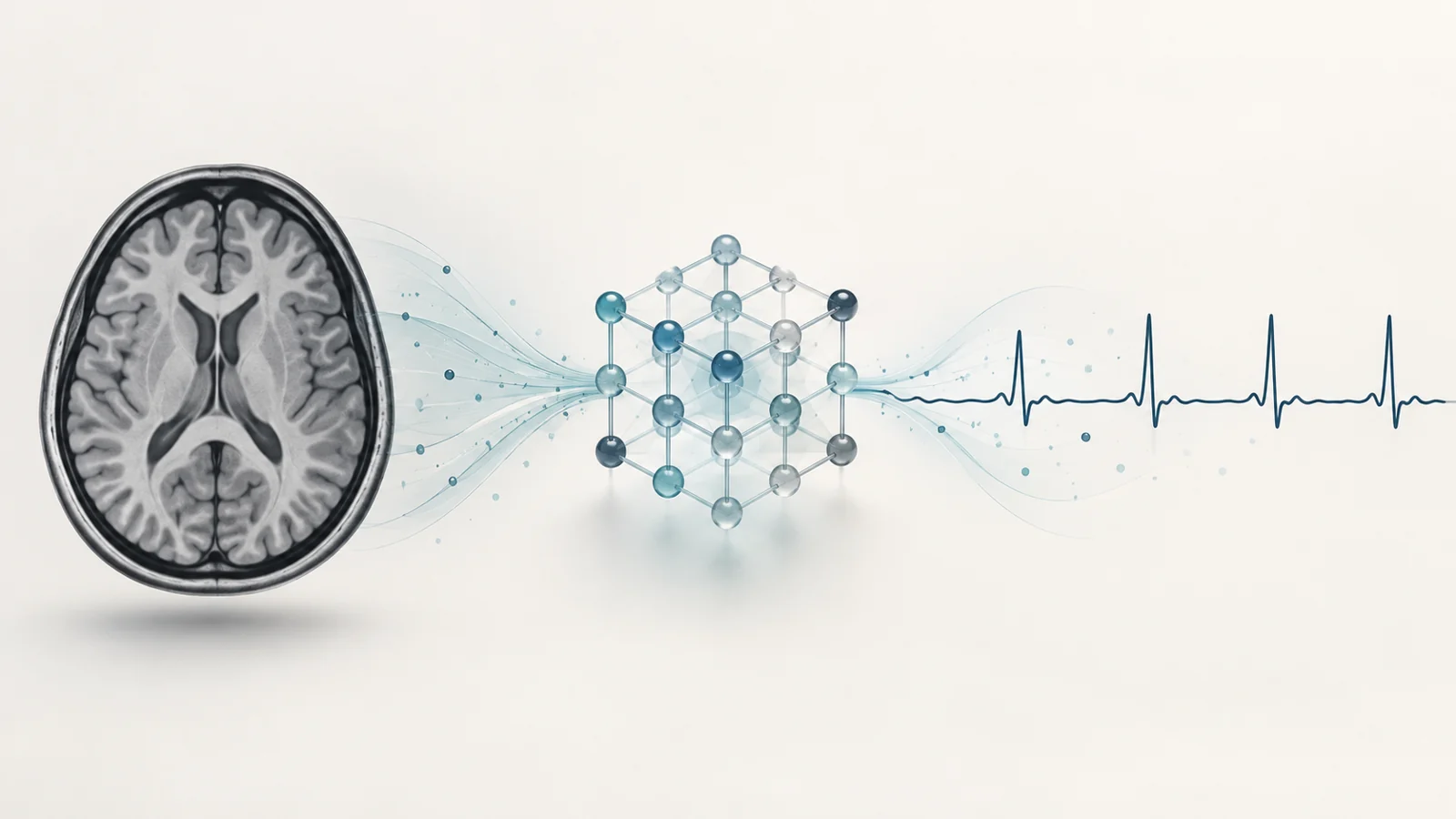
An AI system built to follow the timing of brain activity may have trouble with a still brain scan. That tension runs through a 2025 review of spiking neural networks, which combines a broad survey of neuroimaging applications with the authors’ own quick comparison of models on MRI images.
The difference matters because “brain data” can mean a changing electrical recording or a set of static images. A method that handles one form well may not transfer to the other. The review offers reasons to investigate spiking networks for time-varying data, while its MRI test shows why speed alone is not enough to make them useful.
Learning from the timing of events
Spiking neural networks (SNNs) represent information through discrete events, or spikes, passed between connected units. Their timing carries information: a network can respond to the order and spacing of events rather than treating each input as a steady value. In one learning approach discussed in the review, connections strengthen or weaken according to whether one unit’s spike arrives before or after another’s.
That event-based design is a possible fit for brain recordings that change over time. But it does not automatically give a network a useful way to interpret every kind of image. A static MRI scan contains spatial patterns; it does not arrive as a naturally unfolding series of events. Converting it into spikes requires an encoding choice, and the resulting representation can affect what the model learns.
NeuCube adds a brain-inspired arrangement to the spiking approach. Its three-dimensional structure organizes connections to model spatial as well as temporal patterns. The review describes it as a way to work with multiple data types, including EEG, fMRI, and genetic data, and to combine unsupervised pattern extraction with task-specific learning. This is an architectural proposal for representing relationships in brain data, not evidence that every modality or task will perform equally well.
Faster processing, weaker MRI classification
For its static-image comparison, the paper used a curated subset of the Alzheimer’s Disease Neuroimaging Initiative dataset: grayscale MRI scans resized to 128 by 128 pixels. The set contained 15,888 training images and 2,263 test images across Alzheimer’s disease, normal cognitive status, and mild cognitive impairment. The authors compared a convolutional neural network (CNN), which learns spatial image features, with spiking simulators including SpikingJelly and snnTorch.
The CNN reached 85.9% accuracy. SpikingJelly reached 78.0%, but its precision was 43.5% and recall was 50.0%. Accuracy gives the share of predictions that were correct overall. Precision concerns how often a predicted class was right, while recall concerns how often members of a class were found. These summary figures show weaker classification but do not tell us how the model performed in each of the three groups.
SpikingJelly trained 4.8 times faster and inferred 3.8 times faster than AlexNet, the CNN used in the timing comparison. Those runtime results came with weaker predictive measures. The authors describe the simulator assessment as a quick comparison, without extensive parameter or hyperparameter tuning. It therefore illustrates a tradeoff in this experiment rather than settling how these models would perform after optimization or on other image tasks.
The comparison was made on conventional computing hardware. The authors did not test these models on neuromorphic chips, so the runtime advantage cannot be treated as a measured power saving on specialized hardware. Nor does this curated image task establish diagnostic accuracy or benefit for a broad patient population.
Separate evidence from changing brain activity
The review also recounts prior studies using NeuCube with EEG and fMRI. These involve changing brain activity and different tasks from the static MRI image classification above. The paper describes reported results including EEG classification for epilepsy, migraine, and healthy participants, and analyses of fMRI patterns in cognitive tasks. It also discusses EEG-based motor-imagery work and other brain-computer interface applications.
Those reports help explain why researchers are exploring a three-dimensional spiking model for time-dependent data: the architecture is intended to preserve relationships among activity across brain locations and over time. But the results come from separate studies, with different datasets, tasks, and comparisons. They are not a controlled benchmark against the authors’ MRI experiment, and the review’s account should not be read as independent verification of each cited study.
A useful direction raised by the authors is to divide the work between model types. A CNN could first extract spatial features from an image, while an SNN handles temporal dynamics in subsequent data. The review points to transferring weights from a trained CNN as one possibility for improving SNN performance. This remains a proposed path for further testing, rather than a demonstrated solution in the paper’s MRI comparison.
What still stands between these models and clinical use
Even when a task suits timed data, practical performance depends on how signals are encoded, how the network is trained, and how noisy inputs are handled. The review identifies training complexity, inconsistent methods across SNN systems, integration with established neuroimaging workflows, and limited neuromorphic hardware as obstacles. Noise and variable signal quality can also disrupt the timing patterns on which these models depend.
Clinical translation adds questions about whether a model works across varied patient groups and whether its outputs can be interpreted reliably. The authors call for larger validation across diverse cohorts. Sensitive brain data also raises privacy concerns, while a dataset that does not represent its intended population could produce uneven performance. These issues matter alongside technical accuracy: a fast model is of little use if its errors fall unevenly or its handling of personal data is not trustworthy.
What more responsive brain interfaces could enable
For people with severe motor impairment, more reliable decoding of changing brain activity could eventually support prosthetic control that responds more closely to an individual’s intentions. The review discusses research combining a brain-inspired spiking network with an adaptive classifier to decode motor-related signals. That work points to a possible route toward individually adapted, responsive interfaces, but it does not establish a patient benefit from this review. Reliable performance on noisy recordings and validation across patients would still be necessary before such a capability could be relied on in practice.
Key Sources
- Primary source: Spiking Neural Networks for Multimodal Neuroimaging: A Comprehensive Review of Current Trends and the NeuCube Brain-Inspired Architecture.
DOI or Journal Page · Review or Meta-Analysis · 2025-06-09
Keep exploring