
Research Signal – Source date 2024-06-19
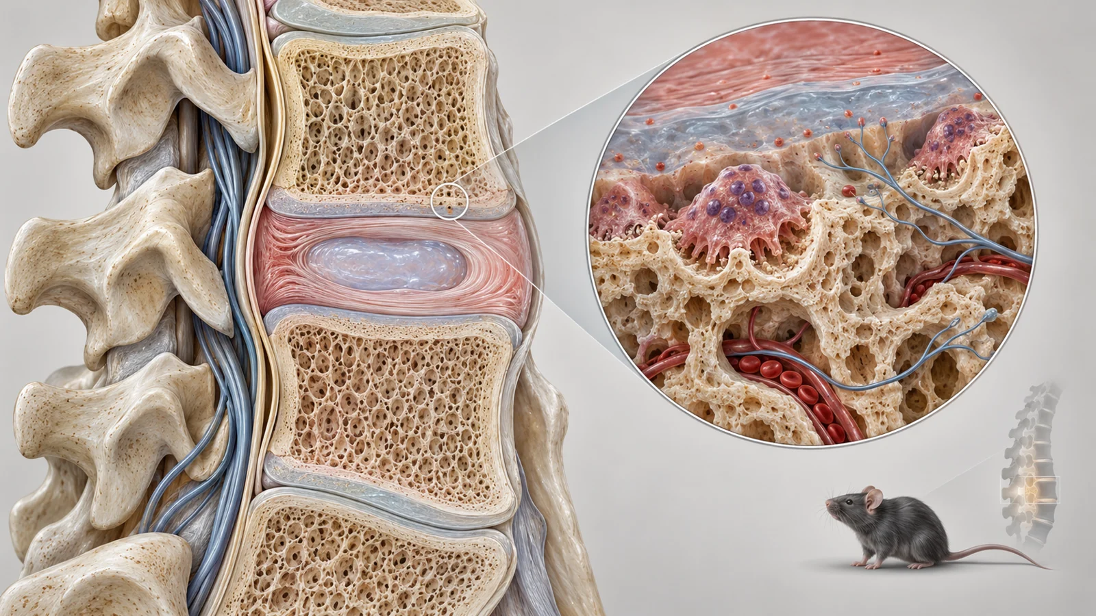
Back pain can arise from several structures in and around the spine. A study published on June 19, 2024, points to one possible contributor: changes in the thin endplates where each spinal disc meets the vertebra above or below it. In mouse models, bone-removing cells in these endplates entered a lasting altered state and were associated with nerve-growth signals, sensory nerve fibers, tissue changes and pain-related behavior.
The researchers then treated the mice with navitoclax, a drug that can eliminate some senescent cells. The treated animals showed fewer of the altered osteoclasts, less endplate porosity, fewer marked sensory nerve fibers and improved behavioral measures. Those results make the endplate a useful place to investigate. They do not show that navitoclax treats human back pain, or that one cell type explains every form of spinal pain.
Where the cells and nerves meet
An endplate is a layer of tissue at the disc–vertebra boundary. It helps connect the disc with the surrounding bone, so changes there can affect the local environment around a degenerating disc. The study examined whether this region could also become more receptive to sensory nerves, which carry information that can contribute to pain sensitivity.
Osteoclasts are part of ordinary bone maintenance. They remove bone as tissue is remodeled, working within a broader process that also includes bone formation. The researchers identified osteoclasts by the TRAP marker and looked for senescence-associated beta-galactosidase and p16, markers used to identify cells in a senescent state. In this context, senescence means that a cell has entered a durable state of growth arrest while remaining biologically active in other ways.
The markers appeared together more often in the endplates of mice with lumbar spine instability and in older mice than in their respective controls. This identifies a population of osteoclasts with senescence-associated features in the affected tissue. It is an important step toward the proposed explanation, but marker-based identification by itself does not prove that these cells caused the pain-related behavior.
Two mouse models test the same idea in different settings
The researchers used two contexts that can produce spinal changes through different routes. In one, three-month-old male mice underwent surgery to create lumbar spine instability by removing the L3–L5 spinous processes and associated ligaments. Sham-operated mice underwent the related procedure without the instability-producing resections. In the other model, 24-month-old male mice represented aging and were compared with younger mice.
These models let the team ask whether the same endplate cell pattern appeared after a structural instability procedure and during aging. In both settings, the affected mice showed changes consistent with spinal hypersensitivity, including more frequent paw withdrawal to mechanical stimulation, shorter heat-response times and less activity on a running wheel. The tests are behavioral measures in mice. They are not direct reports of localized human back pain, and wheel activity can reflect more than one aspect of an animal’s condition.
For the treatment comparison, the researchers gave navitoclax by mouth at 50 milligrams per kilogram per day for seven days, repeated for two cycles with a two-week interval. The full schedule lasted four weeks. The study reports randomization and blinded allocation and outcome assessment. After treatment, the number of osteoclasts carrying senescence-associated markers was reduced in the endplates of the aged and instability models.
What changed after treatment
The behavioral results moved in the same direction in both models. Compared with vehicle-treated mice, navitoclax-treated aged mice withdrew their paws less often in response to mechanical stimulation, took longer to respond to heat and spent more time moving over a greater distance on the wheel. The instability-model mice showed corresponding improvements in paw-withdrawal frequency, heat response, distance traveled and active time.
The study also examined the tissue itself. Microcomputed tomography showed less endplate porosity and less separation between trabecular bone structures after treatment in the tested comparisons. Tissue staining showed lower endplate degeneration scores and reduced distributions of MMP13 and type X collagen, alongside fewer TRAP-positive osteoclasts. These findings describe changes in endplate structure and molecular or cellular markers; they are different from the behavioral measurements and do not demonstrate complete restoration of a healthy spine.
A third set of observations connected the tissue response with sensory innervation. The treated mice had fewer endplate nerve fibers marked by both CGRP, associated with peptidergic pain-sensing fibers, and PGP9.5, a broader nerve-fiber marker. The study also found less growth of marked blood vessels in the endplates after treatment. Vessel growth was a related tissue observation in this experiment, not an independently established cause of the pain-related behavior.
A possible route from bone remodeling to nerve growth
The researchers measured two molecules involved in nerve growth, netrin-1 and nerve growth factor, or NGF. In the aging and instability models, expression of the genes encoding these factors was higher than in the relevant young or sham comparisons. Navitoclax treatment reduced that expression along with the observed reduction in marked sensory nerve fibers.
In plain terms, the proposed sequence is that senescent osteoclasts in a porous, degenerating endplate may help create a local chemical environment that encourages sensory nerve growth. More nerve fibers in an altered tissue could make mechanical or chemical signals easier to transmit as sensitivity. The results support an association between these pieces of the pathway: osteoclasts with senescence-associated markers, nerve-growth-related measurements and sensory nerve staining changed together across the models and treatment groups.
The molecular measurements were made at the endplate level. They show that netrin-1 and NGF-related signals changed in the tissue, but they do not isolate how much came specifically from senescent osteoclasts rather than other affected cells. The authors’ interpretation is supported by the convergence of cell staining, molecular measurements, nerve-fiber staining and behavior, while the experiment does not assign the entire pathway to one cell population.
Why the drug result needs a careful reading
Navitoclax was useful for testing whether removing senescent cells could alter the observed pattern, but it was not a selective tool for senescent endplate osteoclasts. The authors explicitly note that it can affect other cells and has on-target or off-target toxicities. A response after treatment therefore cannot be read as proof that eliminating this one osteoclast population was solely responsible for every improvement.
The same caution applies when moving from mouse behavior to people. Reduced paw withdrawal and increased wheel activity suggest lower hypersensitivity or better activity under the study conditions, but they do not reproduce a person’s experience of back pain. The models also represent particular forms of spinal change rather than the full range of causes behind human back pain. The evidence supplied here does not establish a human therapeutic dose, a durable benefit, or a safe treatment window.
The study’s contribution is narrower and more useful than a claim of a ready-made therapy. It provides an experimental link between senescence-associated osteoclasts in vertebral endplates, signals involved in sensory nerve growth and changes in pain-related behavior in two male-mouse models. It also identifies the central obstacle to translation: a drug that affects senescent cells broadly may not provide the selectivity or tolerability needed for treatment.
What targeting these cells could mean for back pain
If this endplate mechanism is confirmed in people, a future treatment might reduce the cellular signals that encourage painful sensory nerve growth while also limiting some of the surrounding tissue changes associated with degeneration. The potential beneficiaries would be patients whose pain is driven in part by this specific process, rather than everyone with back pain.
Reaching that possibility would require evidence that the same senescent osteoclast and nerve-growth pattern contributes to human endplate-related pain, followed by an intervention that can target the relevant cells sufficiently selectively and safely. From the perspective of this June 2024 study, the result is a plausible therapeutic direction, not clinical availability or proof that broad senolytic treatment will work for people.
Key Sources
- Primary source: Senescence of endplate osteoclasts induces sensory innervation and spinal pain.
DOI or Journal Page · Peer-Reviewed Paper · 2024-06-19
Keep exploring