
Research Signal – Source date 2023-11-13
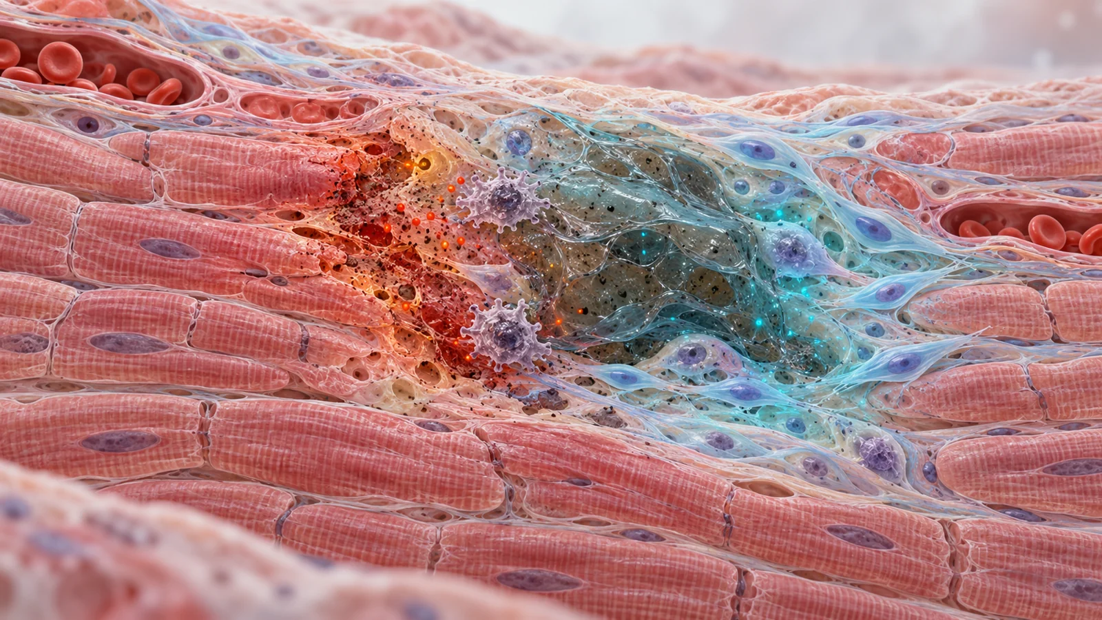
Repairing an aging heart may require more than supplying new cells. The cells also have to survive, remain functional, and work in tissue that has already been damaged. A review published on November 13, 2023, examines how the chemical environment around cardiovascular repair cells may shape that outcome.
The focus is the redox system, the balance between oxygen-related molecules that participate in cell signaling and the defenses that keep those molecules from causing excessive damage. In aging cardiovascular tissue, that balance can shift toward persistent oxidative stress. The result may be weaker stem-cell renewal, cellular senescence, and a less supportive setting for repair.
The review brings together cell experiments, animal injury models, and selected clinical references. Its examples point to a practical idea: regenerative strategies may work better when they protect cells and manage their surroundings. They do not yet amount to a demonstrated way to regenerate damaged human heart tissue.
Repair cells face a hostile starting point
Cell-based cardiovascular treatments have often run into a basic problem. Injecting stem or progenitor cells into a failing heart or injured blood vessel does not guarantee that the cells will remain alive long enough to help. The review cites evidence that a preexisting oxidative environment can kill about 90% of administered cells and impair the function of those that remain.
That loss matters because regenerative treatments depend on a sequence of events. Cells must tolerate the damaged tissue, respond to local signals, and contribute to repair. If most of them disappear soon after delivery, the treatment may have little opportunity to produce a useful effect. Complex biomaterials and delivery systems have been investigated partly to address this survival problem, but the review presents them as developing approaches rather than settled solutions.
Aging adds another layer. Excessive reactive oxygen species, or ROS, can damage DNA, proteins, mitochondria, and other cellular components. Persistent damage is associated with stem-cell exhaustion and reduced regenerative capacity. Yet ROS are not simply waste products to eliminate. At lower levels, they also participate in cell maintenance, differentiation, and reprogramming.
Redox is a timing problem, not a simple enemy
Redox regulation is therefore better understood as a control problem than as a contest between good molecules and bad ones. Cells need enough oxygen-related signaling to respond and change, but too much or too long-lasting an oxidative burden can push them toward dysfunction. The useful range may depend on the cell type, its state, and the stage of a regenerative process.
This is especially important when researchers try to reprogram cells into cardiovascular lineages. The review describes evidence that oxidants can have a time-specific role in reprogramming, while targeted antioxidant intervention may help reduce damaging stress and support later redifferentiation. A blanket instruction to remove all oxidation would miss that timing.
Vitamin C illustrates the difficulty. In one line of work, it improved the efficiency of directly reprogramming mouse fibroblasts into beating cardiac muscle cells. That observation is compatible with redox involvement, but it does not establish that antioxidant activity explains the whole effect. The review also discusses vitamin C’s possible epigenetic action on machinery involved in cellular identity. Other antioxidants, including several that might seem interchangeable, did not reproduce the same effect in forced reprogramming experiments.
The broader lesson is that an intervention can produce a useful cellular outcome without revealing a single mechanism. Dose, concentration, timing, and the specific reprogramming method all matter. Redox-targeting strategies therefore need to preserve the signaling cells require while limiting sustained damage.
Preparing cells before they enter injured tissue
One approach is preconditioning. In plain terms, cells are treated before transplantation so they are better prepared for the environment they are about to encounter. The review describes a concrete example involving human endothelial progenitor cells and an injured mouse artery. Cells preconditioned with a hydrogen-sulfide-releasing compound showed enhanced re-endothelialization, meaning restoration of the vessel’s inner lining, after transplantation.
Hydrogen sulfide is presented as a signaling molecule that can protect against oxidative and inflammatory damage and support adaptive cardiovascular responses. In the mouse artery model, the reported effect was associated with activation of AMPK and endothelial nitric oxide synthase signaling. The experiment therefore connects a preparatory treatment, a specific injury model, and a vessel-repair outcome. It does not show that the same procedure restores aging blood vessels in human patients.
The review also describes another preparation strategy: activating a cell’s own antioxidant defenses. Nrf2 inducers such as sulforaphane reduced intracellular ROS and helped preserve angiogenic behavior in human-derived endothelial colony-forming cells in experimental work. In mouse progenitor cells, another Nrf2 inducer increased antioxidant-gene activity and reduced markers associated with senescence.
These examples shift attention from simply adding cells to improving their readiness. The relevant question is not only whether a cell can be delivered, but whether it can withstand the first encounter with injured tissue and still carry out the function being studied.
Keeping protection close to the injury
A second strategy changes the surroundings rather than the cells alone. Biomaterials can be designed to remain near an injury and provide or carry protective chemistry. Hydrogels are useful in this context because they form a water-rich three-dimensional material that can be placed locally and used to control how associated compounds are released.
The review highlights melanin–alginate hydrogels in a rat model of heart injury. The material was used in the infarct area and was described as scavenging ROS, supporting a more regenerative macrophage response, and suppressing inflammatory and apoptotic responses. The reported experimental outcomes included protection of cardiac muscle cells from oxidative injury, increased angiogenesis, and improvements in cardiac structure and function after the injury.
This is a different intervention sequence from preconditioning. Instead of preparing cells before delivery, the material attempts to make the local tissue environment more tolerable. Other approaches discussed in the review combine biomaterials with antioxidant molecules, Nrf2 inducers, oxygen-related components, or cell and gene delivery systems.
The reported improvements are important, but their meaning needs to stay specific. Better tissue protection, more blood-vessel growth, or improved pumping measures are not automatically proof that new heart muscle formed, integrated with existing tissue, and remained functional over time. A material can influence inflammation and survival without demonstrating the complete process of durable cardiovascular regeneration.
What the experiments can and cannot tell us
The evidence spans several levels, and each answers a different question. Cultured human-derived cells can show whether a treatment changes survival, oxidative stress, angiogenic behavior, or a marker of cellular state. Animal injury models can test how cells or materials behave in a living cardiovascular system. Human studies are needed to examine safety, feasibility, and clinical outcomes in people.
Those levels should not be collapsed into one result. Human origin describes the cells, not the participants in the experiment. A mouse artery model can show restoration of a vessel lining under defined conditions, but it cannot by itself establish reversal of cardiovascular aging. A rat heart-injury model can report improved cardiac function after material placement, but that result does not prove the creation of new, durable heart muscle.
The review’s table also lists IPE-PREVENTION as an ongoing human study focused on circulating endothelial progenitor-cell depletion and dysfunction. That is a different clinical object from demonstrated tissue regeneration. It shows how researchers were investigating the relationship between redox-related processes and circulating repair-cell biology, not that a regenerative treatment had already restored human cardiovascular tissue.
The same caution applies to compounds that appear promising in laboratory settings. The supplied evidence does not authorize a universal antioxidant dose, schedule, or self-treatment recommendation. It supports a research direction in which cellular defenses, delivery materials, timing, and local tissue conditions are studied together.
What better cell survival could mean for heart repair
For older people living with cardiovascular damage, the potential benefit is practical rather than biochemical. A regenerative treatment could eventually help preserve or replace enough functioning tissue to improve the performance of a heart or blood vessel. Better survival would be one step toward that goal because protected cells have a greater chance to remain present, respond to their surroundings, and contribute to repair.
The possibility remains conditional. The cells would still need to produce durable and safe function in patients, and the local environment would need to support their contribution without disrupting necessary signaling. The 2023 review suggests that redox-targeting compounds, protective biomaterials, and more precise cell engineering may help address those bottlenecks. Its evidence supports continued investigation, not a claim that aging hearts can already be regenerated by antioxidants or protected cells.
Key Sources
- Primary source: Targeting the redox system for cardiovascular regeneration in aging.
DOI or Journal Page · Review or Meta-Analysis · 2023-11-13
Keep exploring