
Research Signal – Source date 2022-02-01
Aging bone is shaped not only by the cells that build and remove tissue, but also by the cells that surround them. One possibility raised by a February 1, 2022 review is that some of those surrounding cells enter a persistent stress-altered state called cellular senescence. They change their internal behavior and release inflammatory signals that can influence nearby tissue.
The review brings together cell studies, observations in human bone samples and intervention experiments in mice. Its most direct therapeutic evidence comes from animals: removing senescent cells or suppressing their inflammatory secretions improved several measures of skeletal aging in old mice. That makes senescent cells an important candidate target, but it does not yet show that clearing them prevents fractures or treats osteoporosis in people.
A cellular state that changes bone’s neighborhood
Cellular senescence is not simply a cell becoming old. It is a stress-associated state that can follow signals such as DNA damage or telomere erosion. Senescent cells undergo changes in chromatin and metabolism and become unusually resistant to programmed cell death. In dividing cells, senescence involves growth arrest, but the state can also occur in already nondividing cells such as osteocytes. They also develop what researchers call the senescence-associated secretory phenotype, or SASP.
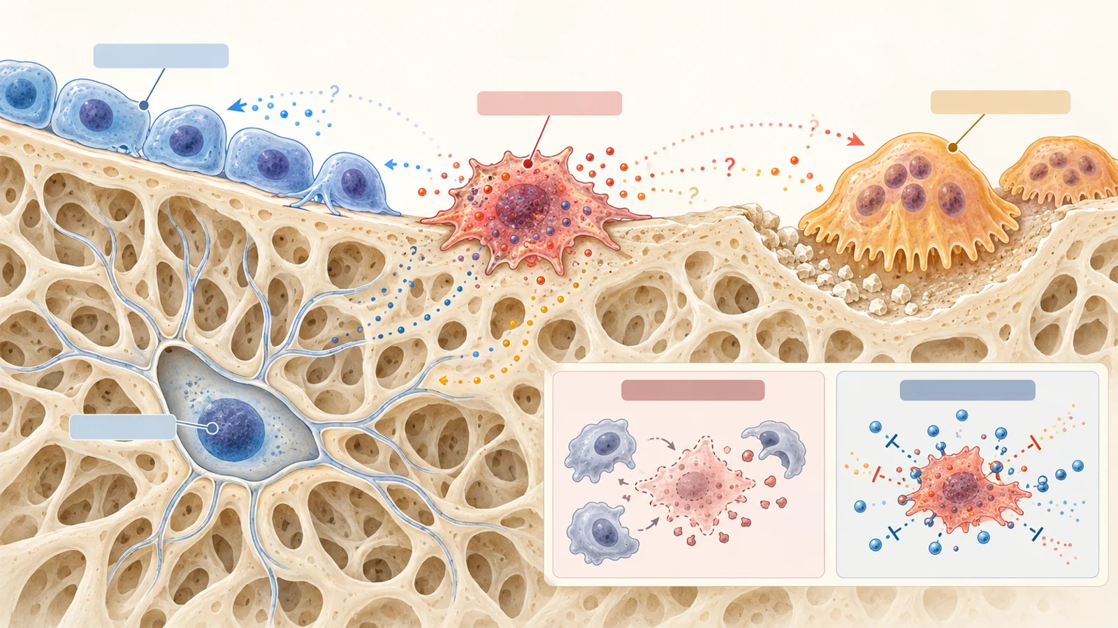
The SASP is a mixture of inflammatory cytokines, chemokines and proteins that can alter the surrounding extracellular matrix. In bone, the concern is less that one senescent cell directly dissolves tissue than that its secretions change the behavior of the cells around it. The review’s working model proposes that a persistent SASP can impair osteoblasts, the cells involved in bone formation; increase osteoclast activity, which contributes to bone resorption; and steer mesenchymal stem cells toward fat cells rather than osteoblasts.
Osteocytes are especially relevant to this question. They are cells embedded within bone. The reviewed work found signs of senescence in osteocytes as well as in osteoblast-lineage cells, mesenchymal stromal cells and immune-cell populations. In older mice, p16Ink4a expression increased across the cell types examined, while p21Cip1 was especially elevated in osteocytes. Bone samples from older women also showed higher p16Ink4a and p21Cip1 expression than samples from younger women.
Those findings support the presence of senescence-associated changes, but identifying a senescent cell requires care. The commonly used SA-β-gal stain is associated with increased lysosomal mass, yet it is not by itself definitive. The review describes a stronger approach that combines several kinds of evidence, including p16Ink4a and p21Cip1, reduced proliferation markers, lower lamin B1, SASP markers and signs of telomeric DNA damage such as SADS or TAFs. This matters because inflammation can resemble part of the SASP without proving that the cells producing it are senescent.
What the aged skeleton reveals
The review’s central biological question is whether senescent cells are merely found in aging bone or help drive its deterioration. The evidence points toward a contribution. In the proposed model, senescent cells accumulate in the bone microenvironment as animals age, and their secretions create conditions that favor less formation, more resorption and a shift in stem-cell fate toward marrow fat.
The model remains a working explanation rather than a complete inventory of causes. The reviewed studies had not established which components of the SASP are most important for osteoblasts, osteoclasts or mesenchymal stem cells. Nor could they determine how much of the effect comes from senescent cells inside bone versus senescent cells elsewhere, such as in fat depots. Signals from the bone environment may also influence distant tissues, but the direction and importance of those exchanges remain open questions.
A striking mouse experiment illustrates why the distinction between local and systemic effects matters. Transplanting a small number of senescent cells into the abdominal cavity of young mice was enough to increase senescence-associated markers in distant tissues, including fat and skeletal muscle, and to produce signs of physical frailty. The cells were not placed in bone, so the result does not identify a skeletal mechanism. It does, however, show how a secretory state in one location might have effects elsewhere.
Removing cells changed bone in aged mice
The most informative tests came from interventions that changed the senescent-cell burden or its secretions. In one genetic model, aged mice carried an inducible mechanism that allowed p16Ink4a-expressing senescent cells to be eliminated. After treatment, the mice had improved trabecular and cortical bone parameters compared with vehicle-treated animals. The changes included fewer osteoclasts, more osteoblasts and higher bone-formation rates on endocortical surfaces.
The response was not identical in every bone compartment. In trabecular bone, formation rates did not rise in the same way, although they were not suppressed as would be expected after reduced resorption and the coupling between osteoclast and osteoblast activity. Bone-marrow adipocytes also decreased in number, perimeter and volume, consistent with a possible shift in mesenchymal stem-cell commitment away from fat and toward the osteoblast lineage.
A second approach used dasatinib plus quercetin, a senolytic combination intended to exploit survival mechanisms that help senescent cells resist apoptosis. A third used ruxolitinib, a JAK inhibitor, to suppress aspects of the SASP without directly removing the cells. In old mice, both approaches produced skeletal effects broadly similar to those seen with genetic clearance. The comparison is useful because it suggests that the harmful influence may involve both the continued presence of senescent cells and the inflammatory signals they release.
It also shows why the findings should not be reduced to a simple “remove one cell type, restore bone” story. The genetic intervention eliminated p16Ink4a-expressing senescent cells throughout the animal. It could not isolate whether the improved bone measurements came from osteocytes, osteoblast-lineage cells, marrow cells, immune cells or senescent populations outside the skeleton. The interventions establish causal leverage in the mouse models, while leaving the responsible populations and molecular mediators less certain.
Estrogen loss points to a context boundary
Postmenopausal estrogen deficiency is another major route to bone loss, but the review does not support treating it as interchangeable with aging-related senescence. In one experiment, the researchers did not find increased expression of the senescence or SASP markers they had used in their aging studies two months after ovariectomy in mice. Clearing senescent cells in young female mice also failed to prevent ovariectomy-induced bone loss or to change the proportion of senescent osteocytes.
Other groups reported findings that pointed in the opposite direction, including increased senescence-associated staining in bone or osteoprogenitor cells after estrogen deficiency. The review discusses several possible reasons for the disagreement. Estrogen deficiency itself creates a proinflammatory state that may resemble some features of a SASP. Some studies relied heavily on SA-β-gal or on antibodies whose specificity was questioned in the review, rather than using multiple markers that can more firmly identify senescence.
Age may also matter. The estrogen-deficiency experiments discussed were performed in relatively young mice, leaving open the possibility that long-term estrogen loss interacts with the separate effects of aging. The practical lesson is not that one result cancels the other. It is that bone loss can arise through different biological routes, and a treatment aimed at senescent cells may not address each route in the same way.
Repair may depend on tissue and timing
Senescent cells are not necessarily harmful in every setting. The review describes a proposed physiological role in tissue repair, where a temporary senescent-cell population may release signals that attract immune cells and activate healing mechanisms. In skin wounds, senescent fibroblasts and endothelial cells have been reported to support repair through secretion of platelet-derived growth factor AA. The proposed sequence depends on the cells being cleared after repair rather than remaining chronically active.
Bone injury adds a different example. In young adult mice, fracture was associated with a temporary accumulation of cells bearing senescence-associated markers in the fracture callus, along with increased p16Ink4a, p21Cip1 and SASP markers. Yet clearing senescent cells with dasatinib plus quercetin did not slow healing. It accelerated the time course of fracture repair in the reviewed experiment.
The contrast does not establish a universal rule about whether senescent cells help or harm healing. It supports tissue-dependent effects; how age, injury setting and timing modify those effects remains uncertain. A therapy that reduces a chronic senescent-cell burden in aging bone may not have the same consequences as an intervention given during an active repair process. That tissue dependence is part of the treatment problem, not a detail that can be ignored once a drug has shown an effect in old mice.
Moving from mouse experiments to human use
The review presents senolytics and senomorphics as early translational possibilities rather than established treatments. As of its February 2022 perspective, human proof-of-concept studies had been initiated, including work directed at age-related bone loss. The review’s argument was that a drug aimed at a fundamental aging mechanism might complement osteoporosis-specific treatments and potentially influence more than one age-related condition.
The route also carries a safety tension. The interventions described in the review generally remove existing senescent cells intermittently or dampen their secretions; they do not permanently prevent cells from ever entering senescence. That distinction matters because senescence can act as an anticancer response to cellular damage. Preventing it broadly could, in principle, remove a barrier to uncontrolled growth. At the same time, senescent cells and their inflammatory secretions may also contribute to tumor-promoting environments, so reducing them could have effects in the opposite direction.
In mice, intermittent administration appeared sufficient to produce benefits in the reviewed work, which could reduce exposure to off-target effects compared with continuous treatment. That observation is a dosing rationale for further investigation, not a safe human schedule. Human studies would need to establish whether the drugs reach the relevant cells, whether skeletal changes are meaningful, which people might benefit and how risks change with repeated treatment. The possibility of improving several conditions at once remains a prospect, not a demonstrated clinical outcome.
What targeting aging could mean for bone health
The larger significance of this research is the possibility of treating weakening bones as one expression of a broader aging process. If future human studies show that carefully selected senotherapeutics can reduce harmful senescent-cell activity without unacceptable effects, older people might gain a way to preserve skeletal health while also improving physical function. The latter could matter for fracture risk because the reviewed mouse work linked senescent-cell clearance with better measures of strength, endurance, walking speed and daily activity.
That combined benefit would differ from treating bone density alone. A therapy acting on a shared aging mechanism could, in principle, address the bone microenvironment and some contributors to frailty at the same time. It could become a complement to osteoporosis care rather than a replacement for every bone-specific treatment. Whether that broader payoff exists in people depends on human evidence showing meaningful benefits and an acceptable balance of harms. The 2022 review makes the idea scientifically plausible; it does not settle the clinical question.
Key Sources
- Primary source: Cellular senescence and the skeleton: pathophysiology and therapeutic implications.
DOI or Journal Page · Review or Meta-Analysis · 2022-02-01
Keep exploring