
Research Signal – Source date 2021-01-01
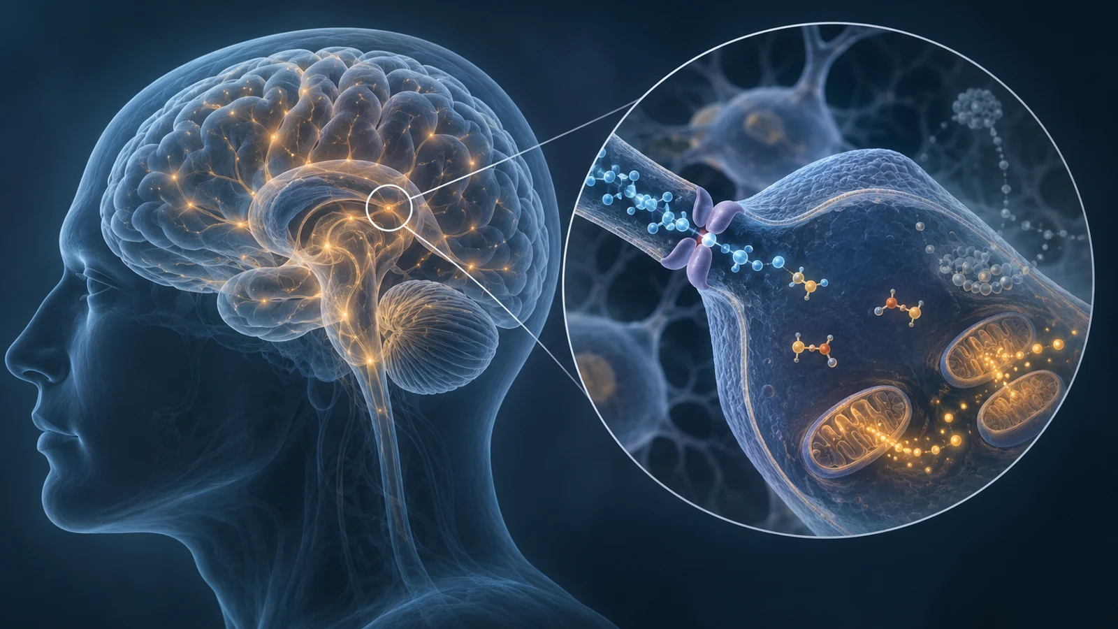
A severe shortage of creatine in the brain sounds as though it should disrupt the machinery that produces energy. Creatine and its phosphorylated form, phosphocreatine, help cells respond quickly when they need more ATP, the molecule used to power cellular work. So what happens when the brain cannot receive enough creatine in the first place?
A study published on January 1, 2021, used magnetic resonance spectroscopy to examine that question in 14 boys with creatine transporter deficiency. The scans showed a profound reduction in creatine-related metabolites. They also showed no significant rise in lactate and pH values within the expected range for brain measurements. The authors interpreted this combination as evidence that oxidative metabolism remained the dominant way the brain supplied energy, even though the creatine-phosphocreatine system was severely depleted.
The finding is informative precisely because it separates two questions that are easy to blend together: how much of the rapid creatine energy buffer is available, and whether the brain’s main energy-producing machinery is still operating in the same broad mode.
The transport problem begins before ATP is made
Creatine transporter deficiency, or CTD, is an inherited disorder caused by pathogenic variants in the X-linked gene SLC6A8. The problem is not simply that the body cannot make creatine. Creatine is produced through steps involving the enzymes AGAT and GAMT, then must cross into the brain and move among brain cells through a transporter. In CTD, that delivery route is impaired.
ATP is the immediate energy currency of cells. Phosphocreatine acts more like a rapidly available reserve: when energy demand rises, creatine kinase can transfer a phosphate group from phosphocreatine to ADP, helping regenerate ATP. This buffering system is especially useful for meeting rapid changes in demand, but it is not the same thing as the whole process of producing ATP.
That difference also explains why results from other creatine deficiency disorders cannot simply be transferred to CTD. In some disorders, the main problem is defective creatine synthesis, and oral creatine can be effective because the transport route remains available. In CTD, most patients respond poorly to creatine supplementation because getting creatine into brain cells is the central obstacle described by the disease mechanism.
What the two spectroscopy methods could see
The researchers used two forms of magnetic resonance spectroscopy. Proton spectroscopy, written as 1H MRS, measured chemical signals including total creatine and lactate. Phosphorus spectroscopy, or 31P MRS, measured phosphorus-containing compounds such as phosphocreatine, inorganic phosphate and ATP-related signals. The phosphorus study was the first application of this method to patients with CTD, according to the authors.
The participants were 14 boys aged 3 to 14, all with genetically confirmed CTD and biochemical confirmation based on their urine creatine-to-creatinine ratio. They underwent the scans under propofol anesthesia. The reported values were compared with healthy measurements published in the scientific literature, rather than with a newly scanned control group in this study.
The proton results showed how large the creatine deficit was relative to a reference metabolite called N-acetylaspartate. The average total creatine-to-NAA ratio was 0.12 ± 0.02 in the participants, compared with published healthy values ranging from 0.65 to 0.81. This ratio demonstrates a markedly weaker creatine signal; it should not be read as an equivalent percentage loss of total brain energy.
The phosphorus results showed a corresponding depletion of phosphocreatine. The tPi-to-PCr ratio was 2.7 ± 0.5 in the acquisition involving all 14 boys and 2.8 ± 0.4 in the longer-interval acquisition involving seven. Published healthy values were 0.26 to 0.44. The PCr-to-γ-ATP ratio was 0.22 ± 0.03 in the first acquisition and 0.26 ± 0.03 in the seven-person acquisition, compared with published healthy values of 1.02 to 1.48.
These are metabolite ratios, not a direct meter of how quickly the brain makes ATP. They establish that the balance of detectable compounds was very different, while leaving the absolute rate of oxidative ATP production and the contribution of each energy pathway unmeasured.
Lactate and pH supplied the study’s key clue
The researchers looked for signs that the brain might be relying more heavily on glycolysis, a route that can produce lactate when energy demand is being met without the same degree of oxidative processing. Lactate was measured at two proton-spectroscopy echo times, including one selected to make the lactate signal easier to assess. No significant lactate elevation was detected at either setting.
The measured intracellular pH was 7.06 ± 0.02, while extracellular pH was 7.51 ± 0.06. The authors reported both values as consistent with normal brain measurements. A lack of elevated lactate together with the pH findings supported their interpretation that oxidative metabolism remained predominant despite the severe reduction in creatine and phosphocreatine.
The wording matters. The scans did not show that mitochondrial function was normal, that energy availability was adequate during demanding cognitive activity, or that the brain had escaped metabolic impairment. They showed a pattern that was more consistent with continued predominance of oxidative metabolism than with a major shift toward glycolytic energy production at rest under the study conditions.
A secondary phosphorus finding added context. In the longer-interval measurements, the researchers detected UDPG, an immediate precursor of glycogen, and found a UDPG-to-NAD ratio of 0.51 ± 0.16 in seven participants. Published healthy values ranged from 0.28 to 0.88. The authors suggested that glycogen-related metabolism may help support oxidative metabolism when the phosphocreatine role is reduced. Detection of a glycogen precursor alone does not demonstrate increased glycogen breakdown or quantify how much energy that pathway supplied.
What these scans leave unresolved
The study offers a detailed biochemical snapshot, but its design places clear limits on how far the interpretation can travel. Fourteen participants is a small cohort, which is expected in a rare inherited disorder but still narrows the precision and generalizability of the findings. All participants were boys between 3 and 14 years old, and the scans were performed under propofol anesthesia. The results therefore do not establish that the same metabolic responses occur in awake children, girls, adults or the wider CTD population.
The longer-interval phosphorus measurements came from only seven of the 14 participants. Comparisons with healthy values also came from published measurements, and differences in participant groups or acquisition conditions can matter when interpreting ratios. The study nevertheless provides a useful first phosphorus-based description of CTD metabolism, especially because it places the very low phosphocreatine signal alongside lactate and pH measurements.
Most importantly, a resting spectroscopy pattern is not a demonstration of performance during speech, learning, movement, seizures or other periods of increased demand. Nor does it show that a proposed intervention will improve development, symptoms or quality of life. The measurements support a physiological interpretation and a therapeutic question; they do not answer the treatment question themselves.
What this could mean for treating creatine transporter deficiency
The authors proposed exploring ways to enhance brain mitochondrial function as a potential treatment direction. For affected children, this could eventually create an additional strategy for supporting energy production when creatine delivery into brain cells remains impaired. The rationale is conditional: mitochondrial support would need to be shown to work safely and to improve meaningful neurological outcomes despite the transport defect.
The 2021 scans did not demonstrate that benefit, and they did not test a mitochondrial enhancer. Their contribution was earlier in the chain. By showing markedly reduced creatine and phosphocreatine alongside lactate and pH findings compatible with continued oxidative metabolism, they suggested that the brain’s energy-producing machinery might be a useful place to look for therapeutic leverage when replacing creatine itself is unlikely to solve the delivery problem.
Key Sources
- Primary source: Oxidative phosphorylation in creatine transporter deficiency.
DOI or Journal Page · Peer-Reviewed Paper · 2021-01-01
Keep exploring