
Research Signal – Source date 2026-03-01
Sleep is often treated as a single health target: more of it must be better. In Alzheimer’s disease, the biology may be more particular. A medicine can make someone sleep without reproducing all of the coordinated chemical and vascular changes that occur during natural sleep.
A review published on March 1, 2026, examines how three wake-promoting neuromodulators—norepinephrine, histamine and orexin—may connect sleep regulation with Alzheimer’s-related protein accumulation. Its central idea is a possible feedback loop: changes in wake-regulating systems could disturb sleep, disturbed sleep could weaken brain fluid clearance, and reduced clearance could contribute to the buildup of proteins associated with Alzheimer’s disease. The review presents this as a proposed relationship assembled from animal studies, human observations, tissue findings and pharmacology experiments. It is not yet a demonstrated causal sequence in people.
Early changes before memory symptoms
Neuromodulatory subcortical systems are collections of deep-brain nuclei that help regulate arousal, attention, cognition and transitions between sleep and wakefulness. They include the locus coeruleus, which produces most brain norepinephrine; the tuberomammillary nucleus, a major source of histamine; and orexin-producing neurons in the lateral hypothalamus.
The reason these systems attract attention in Alzheimer’s research is their timing. The review describes abnormal protein accumulation, neuron loss and altered signaling in several of these nuclei during the preclinical phase, before obvious cognitive decline. It cites evidence that at least 15 neuromodulatory components can show abnormal tau inclusions as early as Braak stage 0, before tau pathology is evident in the entorhinal cortex and before amyloid plaques are established in the usual sequence described for Alzheimer’s pathology.
That ordering raises an important possibility: disruption in systems that coordinate sleep and wakefulness might influence later cortical disease rather than merely appear after memory symptoms begin. But an early biological change is not a personal forecast. The review specifically notes uncertainty about tau in the locus coeruleus. Phosphorylated tau has been observed there even before amyloid deposition in some studies, yet it remains unclear how much of this reflects disease-associated change and how much may be part of aging. Early locus coeruleus tau therefore does not establish that an individual will develop Alzheimer’s disease.
The locus coeruleus is still relevant for another reason. In mice, sleep deprivation has been associated with degeneration of locus coeruleus neurons in a process dependent on norepinephrine. In human observations summarized by the review, locus coeruleus density has shown a strong relationship with later cognitive decline compared with other neuromodulatory nuclei. These findings make the system biologically important, while leaving the direction and timing of cause and consequence unresolved.
Why sleep changes the brain’s fluid environment
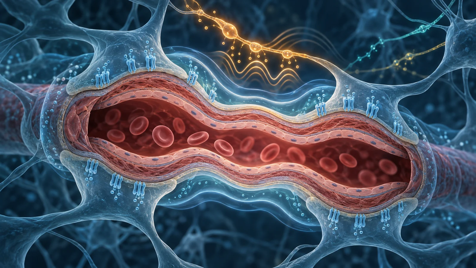
The proposed connection between sleep and protein accumulation depends partly on a physical process often called the glymphatic system. In plain terms, it is a brain fluid-based waste removal pathway. Cerebrospinal fluid moves along spaces around blood vessels and exchanges with fluid between brain cells. Astrocytes, a type of support cell, help organize this movement through water channels called aquaporin-4 at their vascular endfeet.
Arterial pulsations can help drive cerebrospinal fluid into spaces around arteries. The surrounding vascular and astrocyte structures then influence how fluid moves through brain tissue and carries dissolved material away. Meningeal lymphatic vessels provide another route for waste removal, so the review treats glymphatic and meningeal pathways as parts of a broader brain-fluid clearance system.
Sleep appears to be an important state for this exchange, but the mechanism is not simply that every wake signal disappears. Norepinephrine changes vascular tone and can influence the arrangement of aquaporin-4 around vessels. The review discusses evidence that oscillations in locus coeruleus activity during sleep may help coordinate vasomotion, the rhythmic changes in vessel diameter that propel fluid movement around the brain.
That creates a useful resolution to an apparent paradox. Norepinephrine is strongly associated with alertness, yet patterned norepinephrine activity during sleep may still perform a physiological coordination role. A wake-promoting system may need to reduce its sustained alerting drive while retaining carefully timed activity that helps organize vascular and metabolic processes. The relevant question is therefore not only how much sleep a drug produces, but what kind of chemical and physical state accompanies that sleep.
Three wake systems with different vulnerabilities
Norepinephrine, histamine and orexin all support wakefulness, but they do not form one interchangeable pathway. Their receptors, source nuclei and disease associations point in different directions.
Norepinephrine may sit at the intersection of Alzheimer’s pathology, inflammation, vascular behavior and sleep regulation. The review describes a possible sequence in which early beta-2 adrenergic receptor overactivation could increase amyloid production, while rising amyloid later disrupts receptor signaling and reduces noradrenergic support. Receptor loss in Alzheimer’s cortex may affect processes such as microglial surveillance and inflammatory control. This is a model of changing vulnerability over time, not a reason to treat all norepinephrine activity as harmful. The same chemical system may be damaging when persistently dysregulated and useful when operating in coordinated sleep-related patterns.
Histamine has a different profile. Human imaging studies summarized by the review show an age-related decline in H1 receptor binding. Alzheimer’s disease has also been associated with altered histamine levels, degeneration of histamine neurons and increased expression of the H3 receptor in proportion to pathological severity. Because H3 receptors can act as inhibitory autoreceptors, greater H3 signaling could suppress histamine release and influence other neuromodulators regulated through the same network. Animal studies of H3 receptor antagonists have reported neuroprotective effects and better spatial learning, but those findings do not define a proven human treatment strategy.
Orexin, also called hypocretin, is produced in the lateral hypothalamus and projects widely to other wake-regulating nuclei, including the locus coeruleus and histamine-producing tuberomammillary nucleus. It helps initiate and stabilize wakefulness through this broad network. The review links orexin signaling to Alzheimer’s biomarkers, with several cerebrospinal fluid studies reporting higher orexin-A in Alzheimer’s disease or preclinical disease and associations with amyloid and phosphorylated tau. Other studies summarized in the same review did not find differences in orexin-A by amyloid status or cognition. Those inconsistent human associations matter: they prevent orexin from being treated as a uniformly elevated or uniformly causal marker.
The systems may therefore participate in a shared sleep-related problem while requiring different therapeutic logic. Dampening orexin signaling, adjusting histamine feedback or changing adrenergic activity are not versions of the same intervention.
Sleep is not a single pharmacological state
This distinction becomes concrete when comparing medicines that increase sleep drive with medicines that reduce wake drive. Many familiar hypnotics, including zolpidem and benzodiazepines, enhance GABA receptor signaling. The review notes that zolpidem can induce sleep while also inhibiting norepinephrine neurons and abolishing their oscillatory activity during sleep. If those oscillations help coordinate vascular motion, chemically induced sleep could have a different effect on fluid movement than natural sleep.
Dual orexin receptor antagonists, or DORAs, use another strategy. Instead of broadly increasing inhibitory signaling, they block the wake-promoting orexin system. In a randomized crossover study of 51 healthy men summarized by the review, a DORA and zolpidem produced different overnight electrical patterns. Zolpidem increased non-REM sleep while reducing REM sleep and produced large changes in the sleep EEG. Orexin antagonism shortened REM latency, sometimes produced sleep-onset REM episodes and increased REM time without markedly suppressing non-REM sleep. The EEG pattern under orexin antagonism more closely resembled the pattern of natural sleep in that comparison.
That observation is useful for understanding sleep architecture, not for declaring one drug superior for Alzheimer’s disease. A more natural-looking EEG does not by itself demonstrate improved glymphatic clearance, and it does not establish better cognitive outcomes. The comparison does, however, show why drug class matters. Two medicines can both increase time asleep while acting on different neuromodulatory circuits and producing different sleep-stage and EEG patterns.
The review notes that suvorexant, the first approved DORA, has a specific indication for insomnia in Alzheimer’s disease. It also discusses the possibility that orexin antagonism could affect disease biology through more than sleep duration: lower orexin activity might reduce neuronal activity linked to amyloid production and tau phosphorylation, while longer periods of non-REM sleep might support restorative processes. These remain proposed mechanisms rather than established disease modification.
What the evidence can and cannot show
The evidence falls into several different categories, and they should not be treated as equivalent. In mouse models, DORA treatment or orexin gene deletion reduced amyloid plaque accumulation in some APP/PS1 experiments. Other mouse work summarized by the review found no reduction in amyloid pathology, possibly reflecting differences in the aggressive 5xFAD model and the timing of treatment. Lemborexant reduced tau-mediated neurodegeneration in P301S/E4 mice, whereas a similar effect was not observed with zolpidem. These results make orexin biology worth investigating, but animal benefits do not show that all orexin antagonists, disease stages or patient groups will respond in the same way.
Human biomarker evidence is also narrower than a clinical outcome. In healthy participants, a single dose of suvorexant was reported to reduce cerebrospinal fluid amyloid beta and phosphorylated tau, despite a minimal effect on sleep. That is an intriguing acute response. It is not a demonstration that more amyloid was removed from brain tissue, that a clearance pathway accelerated, or that long-term accumulation slowed. A lower fluid concentration can reflect changes in production, transport or other biological processes, and the reviewed finding does not establish prevention of Alzheimer’s disease or slower cognitive decline.
Directly measuring glymphatic function in humans remains difficult. The review describes imaging approaches that infer fluid movement, while noting that many methods remain unvalidated. A proposed wearable approach may offer another approximation, but a dependable, standardized human measure is still needed to connect a change in neuromodulator activity with actual clearance. Without that bridge, sleep stage changes, cerebrospinal fluid biomarkers and inferred fluid dynamics answer related but different questions.
The timing is similarly unsettled. Alzheimer’s disease and sleep affect one another, and the relevant neuromodulatory changes may unfold at different stages. A disrupted system could worsen sleep; poor sleep could then influence fluid dynamics and protein handling; established pathology could further damage the same system. The review’s feedback loop is a useful way to organize these possibilities, but it should not be read as a proven human sequence.
What this could mean for Alzheimer’s treatment
For people at risk of or living with Alzheimer’s disease, the longer-term possibility is a treatment that addresses disturbed sleep while also helping limit the accumulation of pathological proteins. That would be a more meaningful goal than simply adding hours of sedation, because it would connect a person’s nightly brain state with processes that may influence disease progression.
The benefit remains conditional. Targeted changes in norepinephrine, histamine or orexin signaling would need to improve relevant human clearance processes and produce lasting disease benefits, while preserving the waking functions these systems support. The review does not justify treating sleep medicines as interchangeable or starting or stopping any drug. It points instead toward a future in which the chemistry of sleep could be chosen for its effects on the whole brain state: alertness by day, organized sleep at night and, potentially, better handling of Alzheimer’s-related proteins over time.
Key Sources
- Primary source: Wake-promoting neuromodulators in Alzheimer's disease: Implications for sleep and brain clearance.
DOI or Journal Page · Review or Meta-Analysis · 2026-03-01
Keep exploring