
Research Signal – Source date 2023-06-23
Age-related macular degeneration damages the part of the retina responsible for detailed central vision. In its atrophic form, retinal pigment epithelium cells and the underlying choroid gradually degenerate, eventually contributing to geographic atrophy. In neovascular disease, abnormal blood vessels grow into the macular region and can cause more rapid vision loss. Even with available treatments for neovascular disease, long-term visual decline remains a concern.
A review published on June 23, 2023, argues that complement biology is only part of this story. Complement is a protein-based defense system that helps recognize threats, recruit immune cells and coordinate inflammation. Genetic studies have shown that complement regulation matters in macular degeneration, but limited phase 3 evidence for complement inhibition suggests that other processes may also influence disease. The review focuses on a broader possibility: aging may weaken immune regulation while leaving the eye in a persistently inflammatory environment.
A retina that depends on controlled cleanup
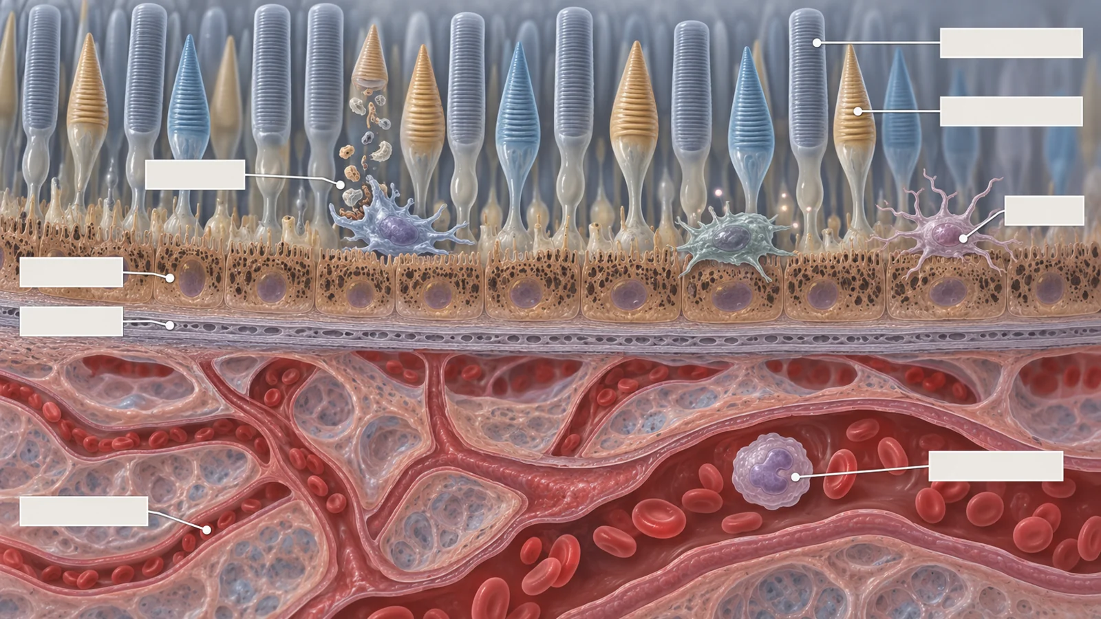
The outer retina is not an isolated sheet of light-sensing cells. Photoreceptors depend on the retinal pigment epithelium, or RPE, a layer that provides structural and metabolic support. Beneath it, the choroid supplies much of the blood needed by the outer retina and contains several immune-cell populations. This arrangement helps explain why changes in both tissue maintenance and local immunity can matter for vision.
The RPE continually handles by-products of photoreceptor renewal. With age, reactive oxygen species, lipofuscin and other compounds such as A2E accumulate around the RPE. These materials can place stress on the cells that support photoreceptors. At the same time, the eye needs some inflammatory activity to identify and clear harmful debris without damaging the surrounding tissue.
The review describes this intermediate state as para-inflammation. It is more active than a completely quiet baseline, but it is intended to preserve tissue balance rather than produce sustained injury. In a healthy setting, low-level immune surveillance can help remove noxious material and resolve local problems. The concern is that aging may disturb the controls that keep this response proportionate.
When protective inflammation loses its balance
Immunosenescence is the age-associated decline in immune regulation and, in some immune cells, in functional performance. It does not mean that every defense simply becomes weaker. Some aging immune cells clear damaged material less effectively, while others produce more inflammatory mediators. The combination can leave unwanted cells or debris in place while increasing the signals that recruit and activate additional immune cells.
This proposed shift is central to the review’s explanation of inflammaging, the chronic, sterile inflammation associated with aging. In the eye, impaired clearance and persistent signaling could disturb the para-inflammatory balance around the RPE, choroid and retina. The model connects systemic immune aging with local tissue stress, but it is an explanatory framework assembled from different kinds of evidence rather than a single demonstrated chain of events in every patient.
The innate immune system is especially important in this discussion. Monocytes, macrophages, neutrophils and natural killer cells help generate inflammatory responses, participate in complement activity, support the blood-retina barrier and communicate with adaptive immune cells. The review describes aging macrophages and neutrophils as having reduced phagocytic activity alongside increased production of inflammatory mediators. Such changes could help explain why cleanup and inflammation can worsen at the same time.
Location changes what immune cells can do
The word macrophage can conceal important differences in location and behavior. Resident macrophages in the choroid can proliferate locally, remove cellular debris and help resolve inflammation. Monocytes from the bone marrow can enter the tissue and develop into infiltrating macrophages with more inflammatory behavior. The review also discusses senescent retinal microglia and circulating immune cells as contributors to a wider inflammatory environment.
These populations are not separated into permanently helpful and harmful categories. The review notes that the familiar laboratory labels for pro-inflammatory and immunoregulatory macrophages are less tidy in living tissue, where cells occupy a spectrum of states. Their contribution depends partly on where they are, what signals they receive and whether they are clearing material or adding to inflammatory stress.
Human observations support an association between immune changes and macular degeneration. The review describes higher peripheral monocyte counts in affected patients, reduced phagocytic function across several monocyte subtypes and higher levels of activated monocytes in some patients with choroidal neovascularization. It also discusses increased IL-17 receptor expression on a subset of circulating monocytes in siblings with macular degeneration. These findings make immune-cell behavior relevant to the disease, but they do not by themselves identify the sequence of events or establish which intervention would help an individual patient.
Complement sits at the intersection of these compartments. Its activation can recruit and activate leukocytes, stimulate inflammatory cytokines and affect the blood-retina barrier. It also helps connect innate and adaptive immunity. Elevated complement fragments have been associated with macular degeneration, and the review describes complement activity as varying with disease stage. The authors’ broader point is that complement may participate in a network of immune communication rather than act as a solitary switch.
Human observations and animal experiments answer different questions
IL-17 provides a useful example of why evidence type matters. The review reports higher IL-17 levels in the blood of patients with macular degeneration and increased expression of part of the IL-17 receptor in macular tissue. Those observations are compatible with a role for IL-17-related signaling in disease, especially because IL-17 can affect monocytes and tissue macrophages.
A separate line of evidence comes from a mouse model, where IL-17 promoted abnormal choroidal vessel growth. The proposed mechanisms included effects on endothelial cells or stimulation of vascular endothelial growth factor production by other cells. This experiment helps show how the pathway might influence neovascular disease in an animal system. It does not establish that the same pathway has the same role in people or that blocking it would improve vision in patients.
The discussion of antibodies illustrates another boundary. Anti-retinal autoantibodies, including antibodies directed toward retinal or extracellular-matrix components, have been described at different stages of macular degeneration. Yet the review characterizes direct evidence for B-cell activity or presence in the choroid as limited. Experimental immune-complex deposition can activate microglia and recruit myeloid cells, but that model does not identify the relevant local process in every human eye.
The result is a layered picture rather than a single immune culprit. Blood measurements may reflect systemic inflammation. Ocular tissue findings may show local responses. Animal experiments can test mechanisms that are difficult to study in people. These forms of evidence can reinforce a biological hypothesis without carrying the same weight for treatment decisions.
Treatment ideas face a targeting problem
The review discusses several ways of addressing immune aging and the inflammatory factors released by senescent cells. A senolytic is intended to remove selected senescent cells. Other approaches aim to reduce the senescence-associated secretory phenotype, or SASP, the collection of inflammatory signals produced by those cells. The distinction matters because clearing a cell and changing what it secretes are different therapeutic actions, with potentially different effects in different tissues.
The review cites studies of dasatinib and quercetin, alone and in combination, as examples of senolytic approaches examined in other clinical contexts. It describes work in diabetic kidney disease in which treatment was associated with reductions in certain senescence-related cellular and circulating inflammatory markers. Those observations concern another disease and do not establish efficacy in macular degeneration.
For the eye, the review discusses UBX1967 as a compound that targeted the anti-apoptotic protein BCL-xL in experimental work on neovascular retinal disease. It also identifies a phase 2 randomized study of UBX1325 in neovascular macular degeneration as ongoing at the time of publication. The study description does not establish an efficacy result, and later outcomes are outside the perspective of this review.
The authors therefore propose that combination or adjunct treatment may eventually be needed. Complement-targeted therapy could address one part of the disease environment while another treatment acts on senescent cells or inflammatory signaling. That remains a treatment hypothesis, not a demonstrated way to prevent progression or preserve sight. It also creates a selection problem: macular degeneration does not progress in the same way for every patient, so broader immune intervention would need to be directed toward people for whom the relevant process is active and the expected benefit justifies the risks.
What immune aging could change about protecting sight
The review’s most important practical possibility is that preserving central vision may require attention to more than one immune pathway. If future research can show that specific features of immune aging and inflammaging are active in suitable patients, and that they can be targeted safely, combination treatment could complement established approaches and reduce the biological pressures that contribute to retinal damage.
That possibility is conditional. The supplied evidence does not show that targeting immune aging prevents macular degeneration or preserves vision in patients. It does suggest why patient selection may matter: treatment aimed at an additional inflammatory process would be more useful if clinicians could identify the people in whom that process is relevant, while avoiding unnecessary intervention for those unlikely to benefit. The larger goal would be concrete and patient-centered—helping suitable patients retain useful central vision as safely as possible.
Key Sources
- Primary source: Beyond the Complement Cascade: Insights into Systemic Immunosenescence and Inflammaging in Age-Related Macular Degeneration and Current Barriers to Treatment.
DOI or Journal Page · Review or Meta-Analysis · 2023-06-23
Keep exploring