
Research Signal – Source date 2024-08-02
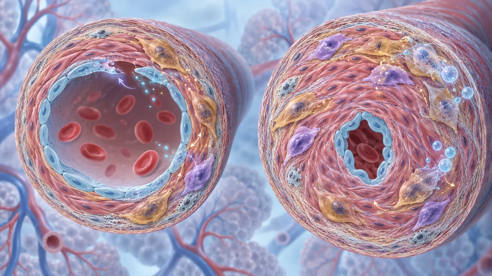
Pulmonary arterial hypertension is a disease in which the small arteries of the lungs narrow or become blocked, raising the pressure that the right side of the heart must pump against. The disease involves injury to the artery lining, inflammation and abnormal growth of the vessel wall.
That is why senescent cells have attracted attention. These are cells that have stopped dividing after stress, but they may remain biologically active. In some settings, their signals help coordinate repair. In others, they can release inflammatory and tissue-remodeling factors that disturb nearby cells. A review published on August 2, 2024, brings together findings that make the therapeutic question unusually difficult: drugs intended to remove senescent cells improved diseased vessels in some animal experiments, yet worsened pulmonary hypertension in others.
The disagreement is not a minor detail around an otherwise settled treatment idea. It raises a more basic question about what these cells are doing at different points in vascular injury, which cells are actually harmful, and whether removing them would also interfere with repair.
Senescence is a state, not a corpse
Cellular senescence is usually described as a lasting halt in cell division. It can be triggered by persistent DNA damage, oxidative stress, mitochondrial problems, oncogene activation or shortened telomeres. The cell does not simply disappear. Instead, it can develop changes in shape and metabolism and release a collection of cytokines, chemokines, growth factors and proteases known as the senescence-associated secretory phenotype, or SASP.
Those secreted factors can alter the tissue around the cell. Senescent cells may support parts of a repair response, while their signals can also promote inflammation or change how neighboring vascular cells grow and move. Senescence is therefore not a single uniform condition with one biological outcome.
Researchers often look for features such as p16, p21, p53 or senescence-associated beta-galactosidase. The review emphasizes a practical problem: no single marker identifies every senescent cell or defines what that cell is doing in a particular artery. A marked cell may be participating in a harmful remodeling process, responding temporarily to injury, or contributing to repair. Finding senescence-associated features in patient tissue is important evidence about what is present, but it is not by itself a reason to remove every marked cell.
When the vessel lining talks to its neighbors
Endothelial cells form the inner lining of blood vessels. They sense blood flow and circulating factors and help regulate vessel tone, permeability and interactions with blood cells. When they are injured, their behavior can change from maintaining a stable barrier to releasing signals that affect the rest of the vessel wall.
The neighboring smooth-muscle cells are one possible target. These cells help give arteries their structure, and changes in their growth or movement can thicken the vessel wall. The review describes an experiment using engineered mice whose endothelial cells were made selectively aged and senescent. Under chronic low-oxygen exposure, these mice developed more severe pulmonary hypertension and greater muscular thickening of the pulmonary arteries than ordinary mice.
The proposed connection was communication between the two cell types. Senescent endothelial cells showed increased expression of Notch ligands, which heightened Notch signaling in nearby smooth-muscle cells. In cell culture, smooth-muscle cells exposed to the senescent endothelial cells became more proliferative and migratory. Blocking Notch signaling with the drug DAPT reduced those effects in culture and reduced hypoxia-induced pulmonary hypertension in the mice.
This experiment supports a mechanism by which endothelial senescence can influence another vascular cell rather than acting in isolation. It does not show that all senescent endothelial cells have the same effect, nor does it reproduce every feature of human pulmonary arterial hypertension. Chronic hypoxia is a useful model of pulmonary hypertension, but it generally produces milder vascular changes than the complex, occlusive lesions seen in advanced PAH.
The treatment schedule changes the interpretation
The clearest disagreement appears in studies of senolytic treatment. Senolytics are drugs intended to trigger the death of senescent cells. One example discussed in the review is ABT-263, also called navitoclax, which blocks BCL-2 family survival pathways.
In one rat model involving congenital heart disease and endothelial injury, senescence-associated markers appeared in remodeled pulmonary arteries as the disease became structurally difficult to reverse. ABT-263 was given after that remodeling had developed. The treatment markedly improved pulmonary hemodynamics and the structure of the vessels. In another rat model using SU5416 and chronic hypoxia, delayed ABT-263 treatment also reduced remodeling and improved hemodynamics in two rat strains.
A different set of experiments reached the opposite result. The review describes pharmacological and genetic interventions targeting senescence in several rodent models. These included ABT-263, the experimental agent FOXO4-DRI, activation of a suicide gene controlled by the p16 promoter, and inactivation of p16 in specially modified mice. These interventions worsened the pressure changes and structural remodeling. The reported adverse findings also included loss of pulmonary endothelial cells.
The schedule was not identical across the studies. In the adverse experiments, interventions began before or soon after pulmonary hypertension was induced. In the same broader research context, ABT-263 given three weeks after monocrotaline-induced pulmonary hypertension produced a modest improvement in pulmonary arterial pressure. The review also notes that the strongest benefit in the congenital-heart-disease model came after occlusive remodeling was already evident.
These observations motivate a repair-versus-remodeling hypothesis. Senescence may be part of an early response that helps preserve or regenerate damaged endothelium, while a later accumulation of senescent cells may help sustain inflammation and obstruction. But the timing pattern is not a proven explanation for every conflicting result, and it does not define a safe treatment window for patients. Differences in species, disease model, drug exposure and the exact cells affected may also matter.
Why animal agreement is not enough
The findings operate at several levels that should not be merged. The review describes senescence-associated features in pulmonary vascular cells from people with PAH and from people with other forms of pulmonary hypertension, as well as in experimental models. Those observations show that the biology is present in diseased vessels. They do not show that a particular cell population is driving the disease or that selectively removing it would help.
The intervention studies are mostly tests in animals. Pulmonary hypertension is a broader category than pulmonary arterial hypertension and can arise in different settings. The chronic-hypoxia model represents one type of pressure and vascular response. Monocrotaline injures pulmonary vascular endothelial cells but also has effects outside the target tissue and usually does not produce the full complex arterial pathology of PAH. The SU5416-plus-hypoxia model produces a more severe phenotype with features that more closely resemble complex pulmonary arterial lesions, but it remains an experimental model.
One possibility raised by the review is that targeting senescent cells can disrupt early repair but reduce harmful remodeling later; the conflicting experiments do not establish that this explains the differences between models. It also matters because a marker such as p16 or p21 identifies a cellular feature, not a complete biological identity. Single-cell and spatial analyses may eventually help determine which endothelial, smooth-muscle or other vascular cells enter senescence, where they are located and how their secreted signals change as disease develops.
The human findings therefore establish a reason to investigate the pathway, not evidence that senescent-cell removal reverses PAH or improves survival in people.
Removing cells is different from quieting their signals
Cell-killing therapies and signal-modifying therapies address different problems. A senolytic such as ABT-263 aims to remove the senescent cell by making it more vulnerable to programmed cell death. A senomorphic, sometimes called a senostatic, aims to change the SASP or block selected inflammatory and remodeling signals without necessarily eliminating the cell.
The second strategy may involve pathways such as NF-kB, p38 MAPK or C/EBP beta, or individual inflammatory factors. It brings its own uncertainty. SASP composition can vary between diseases and over time, and the pathways involved may also perform useful functions in healthy cells. Broadly suppressing those signals throughout the body could therefore affect tissues unrelated to the pulmonary arteries. The review notes that inhaled delivery is one possible way to make lung-directed treatment more specific, but that remains a development challenge rather than an established solution.
A clinical result discussed in the review shows why these approaches should not be treated as interchangeable. Tocilizumab, an antibody that alters inflammatory signaling, did not significantly improve pulmonary vascular resistance in a phase II trial involving people with PAH. That finding does not directly test selective removal of senescent endothelial cells. It does show that a plausible inflammatory target, and benefits from related interventions in animals, do not automatically become an effective PAH treatment in people.
What this could change
The practical payoff would be a more precise way to protect the lung circulation without erasing useful repair. If researchers can identify which vascular cells are persistently driving obstructive remodeling, distinguish them from cells supporting recovery, and confirm a safe target in people, treatment could potentially limit thickening and blockage of the small pulmonary arteries. Preserving more open vessels would help maintain blood flow through the lungs and reduce the workload placed on the right side of the heart.
Reaching that point requires more than showing that a senescence marker is present or that a senolytic works in one rodent model. The relevant cells, their locations, their changing signals and the consequences of treatment would need to be characterized together. Only then could the idea move from an intriguing explanation for conflicting experiments toward a conditional clinical possibility.
Key Sources
- Primary source: Cellular senescence in the pathogenesis of pulmonary arterial hypertension: the good, the bad and the uncertain.
DOI or Journal Page · Review or Meta-Analysis · 2024-08-02
Keep exploring