
Research Signal – Source date 2025-10-27
Endometriosis is a chronic inflammatory condition in which tissue resembling the womb lining grows outside the womb. Lesions commonly occur around pelvic organs and can be associated with pelvic pain, painful periods, pain during sex, painful bowel movements, and infertility. A narrative review published on October 27, 2025, proposes that three biological features may reinforce one another inside this disease: local iron accumulation, persistent cellular senescence, and fibrosis, or scar-like tissue remodeling.
The proposal is not that one simple pathway explains every case. It is an attempt to connect observations that are often studied separately. The review asks whether repeated bleeding can create an iron-rich, oxidative environment; whether some cells then remain permanently stopped in their growth while continuing to send inflammatory signals; and whether those signals help maintain the scarring that makes lesions painful or difficult for surrounding tissues.
A local iron problem begins with bleeding
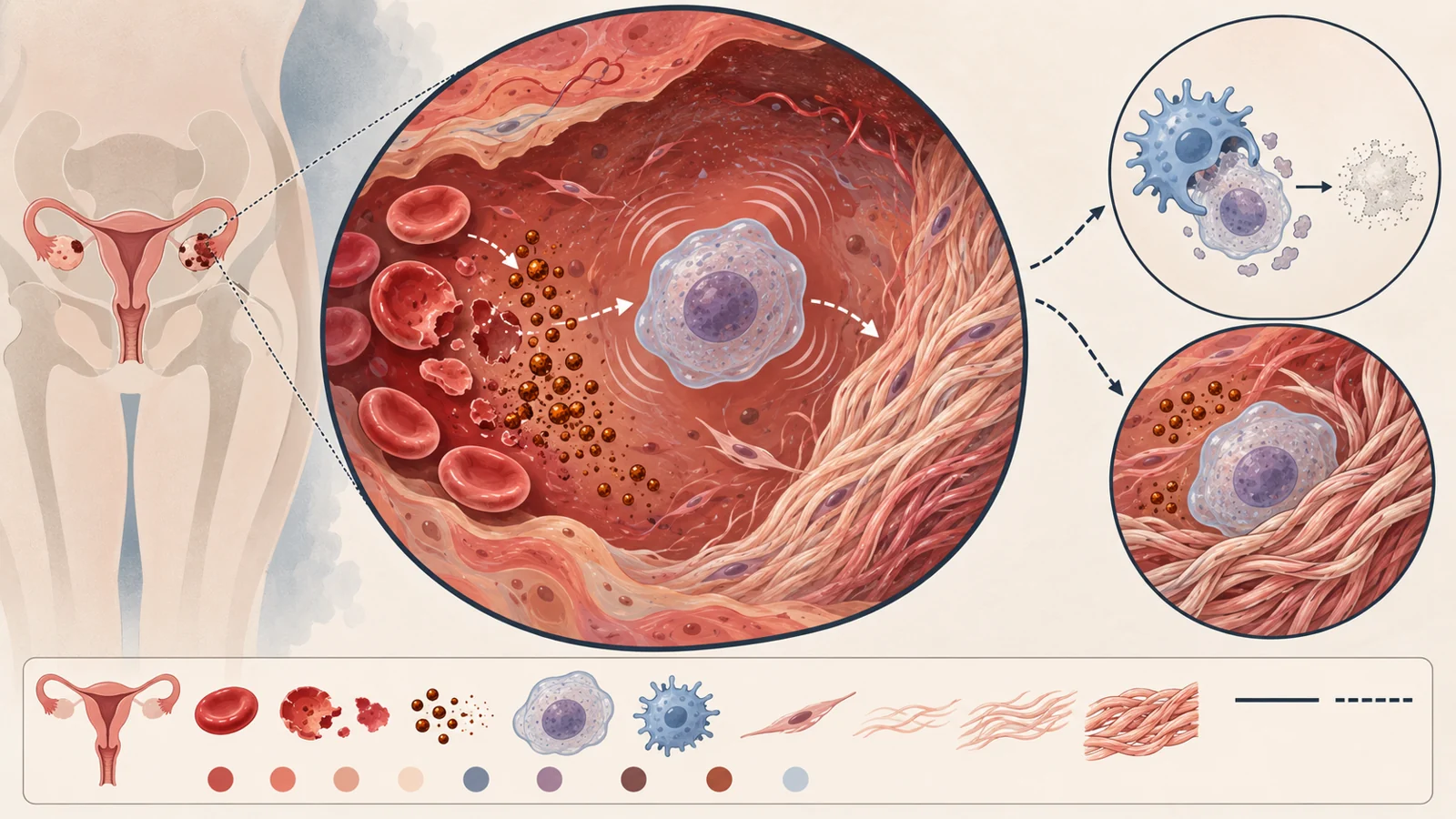
Endometriotic lesions can bleed repeatedly. The review describes blood associated with retrograde menstruation and bleeding within lesions as a likely source of excess iron in the pelvic environment. When red blood cells break down, their hemoglobin is processed into heme and free iron. Peritoneal macrophages normally help remove damaged blood cells and handle their contents, but recurrent bleeding may place more iron into the tissue environment than ordinary storage and transport systems can safely manage.
Several measurements point in the same general direction. Higher free iron, ferritin, and transferrin saturation have been reported in peritoneal fluid, the fluid inside ovarian endometriomas, lesion tissue, and peritoneal macrophages from people with endometriosis. Ferritin is the main storage protein for iron, so increased ferritin can reflect a response to excess iron. Some studies have also found greater iron burden in more extensive disease, although the association has not appeared consistently across all studies.
These findings concern iron around lesions and in local pelvic compartments. They do not show that dietary iron causes endometriosis or provide a reason for an individual to change iron supplements without clinical guidance.
Reactive iron can create oxidative stress
Iron is not automatically damaging. In the bloodstream it is normally carried by transferrin, while cells store much of their iron in ferritin. When transferrin becomes saturated, non-transferrin-bound iron can appear in extracellular fluid. Inside cells, storage can also be exceeded, expanding a loosely bound labile iron pool.
Both forms are chemically reactive. They can participate in reactions that generate reactive oxygen species, molecules capable of stressing proteins, membranes, and DNA. The review presents this shift from safely handled iron to more reactive iron as a possible bridge between repeated hemorrhage and cellular damage in endometriotic tissue.
The important word is possible. Elevated iron measurements and oxidative stress are not, by themselves, proof that iron initiates every lesion or that the same sequence occurs to the same degree in every person. They provide a biological setting in which persistent cell stress could become more likely and deserve closer testing.
A cell can stop dividing without becoming inactive
Cellular senescence is a durable halt in cell division. A senescent cell is not simply dead or inert. It remains metabolically active and can change its shape, internal structures, gene activity, and interactions with nearby cells. Common indicators include increased p16 and p21, activity of SA-beta-galactosidase, loss of Lamin B1, and persistent DNA-damage signals.
The review describes several of these markers in endometriotic lesions, stromal cells derived from lesions, and cells from people with endometriosis. It also discusses higher levels of inflammatory secretions such as interleukin-6 and interleukin-8. These secretions are often grouped under the term senescence-associated secretory phenotype, or SASP. In plain terms, the cell has stopped multiplying but may still release signals that alter the behavior of surrounding tissue.
Presence is not the same as purpose. Detecting senescence markers shows that senescent cells or senescence-like features are present; it does not establish that those cells started a lesion, determine which cells are harmful, or prove that they are the dominant driver of symptoms.
The review proposes that immune surveillance may help explain why these cells persist. Natural killer cells are involved in recognizing misplaced endometrial cells, and they also contribute to the clearance of senescent cells. If natural killer-cell function is impaired in endometriosis, the same immune weakness could permit ectopic cells to survive while also reducing the removal of senescent cells. Persistent cells and their inflammatory secretions would then remain in contact with the lesion and its surroundings for longer.
Scarring changes the tissue around lesions
Fibrosis is the buildup and remodeling of connective tissue, including collagen. In endometriosis, the cells most directly involved are often myofibroblasts. These cells can produce and deposit large amounts of extracellular matrix, especially collagen types I and III. The review identifies transforming growth factor beta as an important regulator of myofibroblast activation and matrix production in this setting.
Repeated inflammation and tissue injury associated with cyclical bleeding could provide continuing signals for this activity. Fibrosis has been observed in superficial peritoneal lesions, ovarian endometriomas, and deep infiltrating endometriosis. As tissue becomes harder and less flexible, scarring can contribute to adhesions and to the way lesions affect nearby structures. The resulting architecture is part of the clinical burden of endometriosis, including pain and impaired reproductive function.
The proposed loop is more than a list of findings
The review’s integrated hypothesis begins with recurrent hemorrhage. Red-cell breakdown increases local iron, and excess reactive iron can promote oxidative stress. Stress may contribute to cellular senescence. Senescent cells may then release inflammatory SASP signals, while incomplete immune clearance allows them to remain. Persistent inflammation and repeated injury can activate myofibroblasts and increase collagen deposition. The review proposes that fibrosis may impair clearance and that recurrent bleeding can replenish local iron. The process could therefore feed back into itself.
This is the review’s proposed iron–senescence–fibrosis cycle, not a fully demonstrated human sequence. Its value is that it gives separate observations a common biological question: whether local iron, persistent senescence, inflammation, and tissue remodeling are connected in time and mechanism. Its risk is that a coherent story can sound more proven than it is.
A plausible mechanism still needs causal testing
The paper identifies itself as a narrative review. Its methods describe searches of PubMed, Google Scholar, and Scopus using terms related to endometriosis, cellular senescence, iron overload, fibrosis, inflammation, uterine aging, and SASP. The authors selected studies for relevance and scientific contribution, with particular interest in mechanistic findings from endometriosis or closely related biological systems.
The evidence comes in different forms, and each answers a different question. Human tissue studies can show that iron markers, senescence-associated features, or fibrosis are present in lesions and nearby compartments. They can also reveal associations with disease features, but association alone cannot establish which event came first. Cell-culture experiments can isolate interactions under controlled conditions, although cells outside the body do not reproduce the full pelvic microenvironment. Animal models can test aspects of a pathway in living tissue, while still differing from human disease. Findings from other tissues may make the proposed biology more plausible, but they are not direct demonstrations in endometriosis.
The review explicitly acknowledges these limits. Human studies are often small and heterogeneous. The connections among iron accumulation, senescence, and fibrosis are sometimes inferred from other systems rather than directly measured together in human endometriotic tissue. Functional experiments will be needed to test whether reducing one part of the proposed cycle changes the others, and whether those changes matter for symptoms or reproductive outcomes.
Treatment would have to separate harmful persistence from useful repair
Senescence is not always harmful. A temporary period of senescence can prevent damaged cells from dividing and can participate in development, tissue remodeling, wound healing, and implantation-related processes. The problem posed by the review is persistent or poorly cleared senescence, not the existence of every senescent cell.
That makes treatment design unusually delicate. Senolytics are approaches intended to remove selected senescent cells. Senomorphics aim to alter harmful secretions without necessarily removing the cells. Other proposals discussed by the review include reducing iron-related toxicity, for example through chelation strategies, or directly limiting fibrotic pathways. These are possible therapeutic directions, not established treatments for pain, fertility, live-birth rates, or existing fibrosis in patients based on the evidence summarized here.
A broad cell-removal strategy could theoretically interfere with useful temporary senescence in the endometrium. A more selective approach might instead focus on chronically persistent cells, particular inflammatory signals, or the local conditions that allow senescence to continue. The review does not establish a patient-selection test, dose, or treatment ranking. It identifies the central design problem: reducing pathological persistence while preserving biology that the reproductive system may need.
What this could change
If the proposed relationships are confirmed in human endometriosis, they could support additional non-hormonal treatment options aimed at limiting lesion-associated scarring and its contribution to pain or impaired reproductive function. The practical benefit would depend on showing that iron-related stress, persistent senescence, or fibrosis can be changed safely and meaningfully in the people who experience them.
Any such approach would need to preserve useful, temporary senescence rather than treating all non-dividing cells as harmful. Validating that balance could eventually make it possible to target the inflammatory and scarring environment around lesions without disrupting processes important for repair and implantation. In October 2025, that remains a promising therapeutic direction and a biological hypothesis requiring further human testing.
Key Sources
- Primary source: The Nexus of Iron, Senescence, and Fibrosis in Endometriosis: A Narrative Review.
DOI or Journal Page · Peer-Reviewed Paper · 2025-10-27
Keep exploring