
Research Signal – Source date 2025-09-05
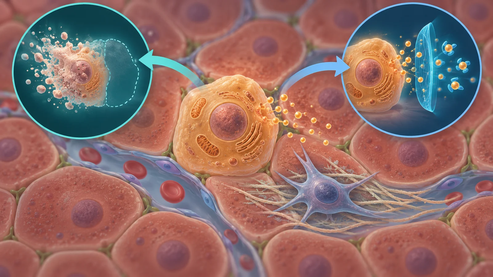
A cell that has stopped dividing may sound inactive. In the liver, that assumption can be misleading. A short-lived senescence response can help contain injury and coordinate repair. When senescent cells persist, their inflammatory signals can instead disturb the surrounding tissue.
That tension is central to a review published on September 5, 2025, on cellular senescence in hepatic disease. The review brings together findings from patient tissue, animal models and cell studies. It does not show that clearing senescent cells improves liver disease in people. Instead, it helps explain why a seemingly simple treatment idea—remove the damaged cells—depends on which cells are involved, when they became senescent and what they are doing in the tissue.
A useful stop that can become a problem
Cellular senescence is a lasting halt in cell division triggered by stress or damage. The cell does not simply disappear, though. It can remain metabolically active and change its shape, while releasing a mixture of cytokines, chemokines, growth factors, oxidative factors and enzymes that remodel the surrounding material. Researchers call this mixture the senescence-associated secretory phenotype, or SASP.
A short-lived senescent response can be part of normal biology. During an acute injury, senescent cells may help recruit and activate immune cells, supporting tissue repair and limiting further damage. The problem changes when senescent cells persist and accumulate. Their secretions can maintain inflammation, alter the local tissue environment and encourage neighboring cells to adopt senescence-related behavior. Signals can act on nearby cells or travel farther through the tissue, so a small population may have effects larger than its numbers suggest.
The review describes this as a balance rather than a universal rule. A tissue may tolerate a limited burden of senescent cells, while chronic injury, aging or impaired clearance allows the burden to rise beyond that tolerance. At that point, continuing secretion can contribute to inflammation and abnormal tissue remodeling. Removing every cell that carries a senescence marker would therefore risk confusing a protective temporary response with a harmful persistent one.
The cell has stopped dividing, not communicating
The liver contains several cell populations that respond differently to injury. That matters because senescence is not one uniform event across the organ.
Hepatic stellate cells provide a useful example. They are normally quiet, but injury can activate them into several functional states, including states associated with inflammation or scar formation. The review discusses evidence that a subset of activated stellate cells contributes to the inflammatory and fibrotic effects associated with SASP. Transforming growth factor beta is highlighted as a major regulator of stellate-cell activation and of their transition toward collagen-producing myofibroblast-like cells.
The biology is not reducible to “senescence is good” or “senescence is bad.” The review also describes a setting in which continued stellate-cell proliferation, together with impaired senescence mechanisms, creates a permissive environment for fibrosis and potentially for liver cancer development. In that context, limiting harmful signaling might be useful, but the fact that growth arrest can occur in one population does not by itself mean that eliminating those cells will reverse scarring. Growth arrest, inflammatory secretion and extracellular-matrix production are related processes, not interchangeable measurements.
Fatty liver findings answer different questions
The review uses metabolic-associated fatty liver disease, or MAFLD, to connect observations in people with experiments in animals. The review reports more senescent hepatocytes in people with MAFLD and a biopsy-based association between hepatocyte senescence and disease severity. It separately reports elevated senescence markers, including SA-β-Gal, p16, p21 and p53, in patients with fatty liver disease.
That observation is important because it links senescence-related features with the severity of a human liver condition. It does not, by itself, establish that senescence initiated the disease. A damaged or inflamed liver may create conditions that promote senescence, while senescent cells may then intensify the damage. The biopsy finding also does not show that changing one of the markers would improve how a patient feels, how the liver functions or how the disease progresses.
Rodent experiments ask a different question. In high-fat-diet models, changes in pathways involving p16, p21, cell-cycle control and oxidative stress accompany hepatocyte arrest and fatty-liver disease. Other mouse experiments have reported changes in lipid handling, cell damage or disease features after manipulating senescence-related pathways or clearing cells that express particular markers.
Those interventions are more informative about possible causation than an association in a biopsy, but they remain model-specific. A mouse result does not establish one treatment across fatty liver disease, viral hepatitis, alcohol-related disease, cirrhosis and liver cancer. Nor does a reduction in a senescence marker automatically translate into a meaningful benefit for a person. The biological question is not only whether a marker falls, but whether the intervention improves liver function or reduces clinically important progression without disrupting repair.
Two ways to target senescence
The therapeutic approaches discussed in the review fall into two broad categories. Senolytics are intended to eliminate senescent cells by exploiting survival pathways that help those cells resist death. Senomorphics leave the cells in place but aim to reduce damaging SASP activity through pathways involved in inflammatory and metabolic regulation.
Dasatinib plus quercetin illustrates the senolytic idea in mouse liver models. In aged mice, the combination reduced senescence-associated features and cleared p16-positive liver cells. Separate high-fat-diet experiments reported reduced fatty-liver pathology. In another mouse model involving altered hepatic metabolism, intermittent treatment was associated with removal of senescent stellate cells and slower progression of liver-cancer-related changes. The outcomes here are experimental measurements in mice, not evidence of a clinical treatment effect.
The same review also describes a cautionary result: another preclinical study found that the combination worsened liver pathology and tumorigenesis without effectively clearing the targeted cells. That inconsistency is not a minor footnote. It shows why a drug’s label as “senolytic” does not guarantee the same biological result in every liver condition or cell population.
Nicotinamide, an NAD+ precursor, represents a senomorphic strategy in the review’s discussion of alcohol-related liver injury. In mice, it was associated with improved metabolic function, lower liver-injury markers and reduced SASP production while acting through pathways connected with cellular senescence. The measured outcomes were changes in the animals’ metabolism, injury markers and inflammatory signaling. They were not patient outcomes, and the example does not establish an appropriate human dose or schedule.
The hard step is showing a patient benefit
Moving from these experiments to a usable liver treatment requires more than finding a compound that changes senescence biology. Researchers would need to know which disease is being treated, which liver cell population is driving harm, whether the target is the senescent cell itself or its secretions, and whether the intervention leaves beneficial repair intact.
The review emphasizes that liver-directed senescence interventions remain largely preclinical, based mainly on animal and laboratory studies. Questions about efficacy and safety in patients remain open. So do practical treatment parameters such as dose, route of administration and whether combinations are appropriate. A strategy that affects several survival or inflammatory pathways may behave differently in a person with one form of liver disease than in a carefully selected mouse model.
Patient observations still have a valuable role. They can show that senescence-related markers and cell states track with disease severity, helping identify biological relationships worth testing. Animal and cell experiments can then probe whether changing those relationships alters disease features. The evidence becomes clinically persuasive only when an intervention produces a meaningful benefit for patients while maintaining an acceptable safety profile.
What this could change
Targeting persistent senescent-cell signaling could eventually help people with chronic inflammatory liver disease retain more functioning liver tissue and face less progression toward scarring. That possibility depends on interventions producing a real clinical benefit safely in the particular disease and cell context, rather than merely lowering a marker or clearing cells indiscriminately. The review’s contribution is to make the treatment problem more precise: preserving useful repair may require controlling harmful signaling, selecting the right cell population and proving that the change matters to patients.
Key Sources
- Primary source: Role of cellular senescence in hepatic diseases (Review).
DOI or Journal Page · Peer-Reviewed Paper · 2025-09-05
Keep exploring