
Research Signal – Source date 2023-06-08
An implant designed to disappear has a demanding job. It must give damaged tissue somewhere to rebuild, then relinquish that role as living tissue develops. If its structure breaks down before the replacement can provide support, regeneration may be undermined. Remaining in place longer also has consequences, including continued interaction with the immune system.
These temporary supports are called resorbable scaffolds, materials designed to break down and be absorbed by the body. A review published on June 8, 2023, examines the polymers and material combinations used to construct them, along with the body’s response after implantation. It brings together research across hard and soft tissues rather than reporting a newly tested implant.
The central challenge extends beyond finding a material that cells tolerate. A scaffold has to provide accessible space, suitable surfaces and useful mechanical properties in a biological environment that changes around it. Understanding those jobs helps explain why encouraging laboratory observations are only part of the evidence needed for tissue repair.
A support with room for living tissue
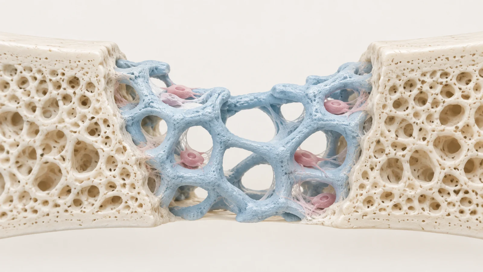
Picture a three-dimensional network of fibers or struts with connected spaces between them. Cells need access to the interior, where they can attach, multiply and deposit extracellular matrix—the material surrounding cells that helps give tissue its structure. Oxygen and nutrients must reach those cells, while metabolic waste must leave. A scaffold’s empty spaces are therefore part of its function.
The arrangement matters as much as the presence of holes. The review identifies pore distribution, exposed surface area and overall architecture as features that influence cell penetration and matrix formation. Surface chemistry also affects whether cells can attach. A structure can look suitably porous while still presenting surfaces that are difficult for cells to occupy.
Opening up more space introduces a physical tradeoff. Larger pores can favor biological responses associated with regeneration in studies discussed by the authors, but too many large openings can weaken the structure. Bone-oriented scaffolds must meet different mechanical demands from supports intended for skin or other soft tissues. These differences prevent a single pore size or architecture from serving as a universal design.
What combining materials makes possible
Collagen illustrates why a biologically familiar ingredient may need reinforcement. The review describes collagen scaffolds used to construct engineered skin as often having poor mechanical properties. Combining collagen with polycaprolactone, or PCL, offers a way to strengthen the structure. The mixture can be formed into fibers through electrospinning, a fabrication method discussed for skin and vascular tissue engineering.
Here, blending lets researchers construct a fibrous support that brings collagen together with the mechanical contribution of another polymer. The useful question is how that particular combination behaves as a scaffold, including its surface properties and strength.
For bone, the ingredients and construction goals differ. The review discusses combining polylactic acid, or PLA, with bioceramics such as hydroxyapatite. These composites bring a polymer together with a mineral component studied for bone regeneration. In the examples reviewed, combining the materials can improve bending and compression performance and make fracture behavior less brittle than that of the ceramic alone.
Some PLA–hydroxyapatite combinations can also be processed by fused deposition modeling, a form of 3D printing. That creates a route toward making porous structures in customized shapes. The processing advantage is concrete: the material combination helps determine what engineers can manufacture, as well as how the finished object behaves.
The labels applied to these ingredients require care. Naturally occurring polymers, including collagen and chitosan, are different from polymers synthesized using biomass-derived building blocks. PLA is an example of the latter. “Bio-based” describes the origin of its feedstock; it does not mean the finished polymer occurs naturally. Neither label establishes freedom from inflammation, toxicity or manufacturing variability. Composition and the properties of the finished scaffold remain essential.
How water and chemistry control breakdown
For several polymers discussed in the review, breakdown involves hydrolysis. Water breaks susceptible chemical bonds in the material. The chemistry of those bonds, the polymer’s composition and the way its chains are connected give researchers ways to influence decomposition.
Poly(propylene fumarate), or PPF, provides a specific example. Its chains can be crosslinked into a network that contributes mechanical strength. The review explains that its hydrolytic degradation depends on the molecular mass of the chains, the crosslinking agents and the density of those connections. Changing the network therefore changes more than the initial strength of the implant; it can also alter how the material breaks down.
Blending offers another set of controls. PLA has limitations that include poor wettability, difficulty supporting cell attachment and slow hydrolytic degradation. Combining it with other materials can modify these properties. The authors also describe mineral additions that alter degradation in PLA/PCL combinations. Such findings concern particular formulations, rather than a rule that any added ceramic or polymer will have the same effect.
These controls address the opening timing problem. Researchers want the loss of scaffold support to correspond to the formation of replacement tissue. But a tunable material is not an exact biological clock. A useful formulation has to be assessed for the intended tissue and structure; the review does not establish a resorption schedule that can be transferred across applications.
Why the body may wall off an implant
Implantation sets inflammatory and wound-healing responses in motion. The eventual result can include tissue regeneration, but it can also include fibrosis, in which the body forms a fibrous capsule around the foreign material. That enclosure is a different outcome from cells integrating the scaffold into rebuilding tissue.
Macrophages, immune cells involved in these responses, appear throughout the review’s discussion of scaffold design. Studies it describes link fiber diameter and alignment, pore dimensions, stiffness and surface chemistry to changes in immune behavior. Some findings associate larger, aligned fibers with macrophage characteristics linked to repair. These observations suggest that physical architecture can influence biology as well as provide support.
Animal findings reported by the review also show why material origin is an incomplete guide. In one comparison, biological scaffolds elicited immune features associated with regeneration, while fossil-based scaffold implants showed features of chronic inflammation. Yet all implanted groups had greater immune responses than untreated animals. Those results describe the tested materials and conditions; they cannot make every naturally derived scaffold a safer choice.
Breakdown products add another influence. Acidic products released as some materials degrade can intensify local inflammation and favor fibrosis. The authors discuss incorporating basic substances to counter excess acidity as a possible design response. Degradation is thus a chemical event experienced by surrounding cells, with consequences beyond the gradual loss of the scaffold itself.
Cell growth is one result, restored function is another
The evidence assembled in the review answers several different questions. Mechanical testing measures properties such as resistance to compression or bending. Cell experiments examine attachment, spreading and proliferation. Animal observations reveal aspects of inflammation and tissue interaction within a living host. Each contributes useful information, but the endpoints are not interchangeable.
For example, the review describes PLA structures coated or filled with collagen that supported the growth of several cell types, including bone-related cells and cells from human umbilical vein endothelium. That is evidence about cellular compatibility under the conditions tested. It does not by itself establish that an implanted structure will restore durable tissue function in a patient.
Likewise, a favorable immune marker cannot stand in for a functioning replacement tissue. The findings span different formulations, fabrication methods and anatomical uses, so they do not form a common head-to-head contest from which one best scaffold can be selected.
Mechanical durability deserves particular attention. A structure that performs well in a strength test may face repeated loading in the body. The authors identify fatigue limits under recurring stress as an area requiring clearer understanding. For a bone application, the relevant question includes how the construct performs through those repeated demands, alongside its interaction with developing tissue.
The review also notes established surgical uses of resorbable polymers, including fixation materials and sutures. Those uses provide context for the materials, but they do not confer clinical effectiveness or regulatory approval on every porous scaffold containing them. The authors explicitly identify formulation-specific optimization and translation into clinical practice as continuing work.
What this could change
For patients with bone defects, one prospect is a graft shaped to the damaged region that provides temporary support and guides new bone formation. The causal path combines several capabilities described in the review: printable polymer–ceramic mixtures, control over the scaffold’s porous geometry and material properties that encourage interaction with surrounding tissue.
If manufacturing, tissue integration and sustained mechanical performance can be validated together, such grafts could make the shape and internal structure of a support more closely suited to a particular defect. A correctly shaped object would then become useful through its biological and mechanical performance after implantation.
The review also discusses in situ tissue engineering, in which a bioactive scaffold is implanted without added cells and is intended to recruit the body’s own healing response. Applied successfully to a bone defect, that approach could help living tissue develop at the repair site without first constructing a cell-seeded replacement outside the body. From the June 2023 perspective, patient-shaped regenerative grafts remain a conditional clinical prospect, supported by useful material and fabrication research whose combined performance still needs validation.
Key Sources
- Primary source: Resorbable Biomaterials Used for 3D Scaffolds in Tissue Engineering: A Review.
DOI or Journal Page · Peer-Reviewed Paper · 2023-06-08
Keep exploring