
Research Signal – Source date 2023-04-03
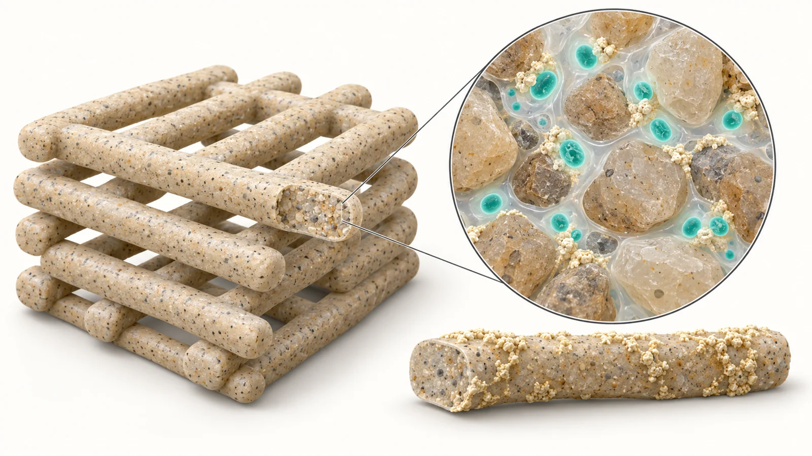
The object is small enough to sit on a fingertip: a lattice of gel-and-sand strands, deposited through a printer nozzle with open gaps between them. Inside the strands are living photosynthetic bacteria. Given light and a suitable liquid medium, these cells can participate in forming calcium carbonate, a mineral that could help bind the material together.
In a study published on April 3, 2023, researchers combined this mineral-forming capacity with extrusion-based 3D bioprinting. They made laboratory specimens containing the cyanobacterium Synechococcus sp. strain PCC 7002 and compared them with matching specimens made without cells. The bacteria survived printing, and the resulting materials behaved differently under compression. One dried, sand-rich formulation resisted compression much more strongly with cells; another became weaker.
That split result is central to understanding what was made. The experiment brings together a way to shape a material and a possible way to develop mineral binder inside it. How those parts interact depends on the mixture, its condition during testing, and exactly what the test measures.
Getting a living mixture through a nozzle
The printable paste combined alginate and methylcellulose, two gel-forming ingredients, with fine sea sand and a suspension of bacteria. The gel carried both the cells and the grains through the nozzle. After printing, a calcium chloride treatment crosslinked the alginate, helping the deposited structure hold together.
A useful printing paste must flow under pressure while retaining its shape after deposition. These mixtures were shear-thinning, meaning that they became less resistant to flow as they were pushed through the nozzle. Adding sand increased viscosity at low shear rates, but the mixtures flowed similarly under the higher shear conditions tested. Sand also improved shape retention in the printed patterns.
There was a processing limit. Mixtures containing up to 50% sand by weight formed a homogeneous, printable paste. The tested 70% mixture was too stiff and brittle for extrusion bioprinting. Increasing the amount of solid filler therefore could not simply continue until the material resembled a conventional sand-rich building product.
Printing offered control over the spaces as well as the solid strands. The mineralization specimens were roughly centimeter-sized lattices, with successive layers running across one another. Their open gaps are potentially useful because living cells need access to their surroundings. A designed pore structure could make it easier to deliver light and medium through a material, although the geometry that supports cultivation also has to serve the component’s eventual mechanical purpose.
What survived the printing process
The researchers checked whether extrusion pressure and contact with sand damaged the bacteria. Fluorescence microscopy used chlorophyll’s natural red glow to identify living cells, alongside a dye that marked dead cells. Separate chlorophyll measurements tracked changes in the material’s photosynthetic biomass.
In the general cultivation experiment, measured viability on the first day was about 87–93% across the tested formulations. Chlorophyll content increased during the first week, supporting the finding that cells remained biologically functional after immobilization in the gel.
The observations extended through 14 days, but the cells did not all remain neatly confined. During prolonged cultivation, bacteria grew out into the surrounding medium, particularly from sand-containing specimens. The authors suggested that soluble methylcellulose washing out, together with spaces around sand grains, could open routes through the gel. By day 14, measured viability in the 50% sand formulation was lower than in the sand-free gel.
The mineralization experiment was a separate assessment under different medium conditions. Here, viability after 14 days was approximately 74% in sand-free specimens and 81% in specimens containing 30% sand. This establishes persistence during the tested cultivation period. It does not extend the survival finding to dried specimens or to a finished component exposed to years of changing moisture, temperature, and light.
Minerals appeared without bacteria too
To encourage mineral formation, the researchers supplied a medium containing calcium chloride and sodium bicarbonate. The specimens were cultivated under controlled light and temperature. This chemical environment matters to the interpretation because it could also produce mineral deposits without living cells.
Electron microscopy showed mineral-covered surfaces on both the living specimens and the cell-free controls. A mineral crust on the outside was therefore insufficient evidence of a bacterial contribution. The surrounding medium was supersaturated, and the scaffold surface provided places where precipitation could begin.
The more informative comparison lay inside the printed strands. In examined cross sections containing 50% sand, the cell-free material had a relatively smooth gel matrix between grains, with few visible mineral deposits. The material containing bacteria had many more internal deposits, including deposits around cells and along the matrix between sand grains. Elemental analysis detected calcium, consistent with the authors’ identification of calcium carbonate in this experimental setting.
Together, these observations support a cell-associated contribution to mineral formation within the material. They also suggest a possible route for consolidating sand grains through mineral deposition. The experiment does not fully separate active biological effects from passive effects of the cells themselves. The authors identified a dead or inactivated-cell control as a way to investigate that distinction.
The strength result changes with the test
The mechanical findings require two separate comparisons. Fresh, wet scaffolds were tested after removal from the mineralization medium. A second set was cultivated for seven days and then air-dried to stable weight before testing. The reported endpoints also differed.
For the wet scaffolds, the researchers compared maximum compressive strength. Cell-containing specimens exceeded their matching cell-free controls at each measured time point—one, seven, and 14 days. After one day, the increase was about 38% in the sand-free formulation and 24% in the formulation containing 30% sand.
Neither the living specimens nor the controls showed a significant increase in maximum compressive strength over that cultivation period. Continued time in the medium did not produce a steadily rising strength result under these conditions. The authors suggested that mineral formation might have reached an early equilibrium; renewing the medium was one possible avenue they discussed for changing the process.
For the dried specimens, the comparison was compressive stress at 50% strain. That means the stress measured when a specimen had been compressed to half its original height. It is a large-deformation endpoint, and it needs to stay attached to the numbers.
| Sand content by weight | Cell-free dried specimens | Dried specimens containing PCC 7002 |
|---|---|---|
| 30% | 25.7 ± 2.4 MPa | 14.4 ± 1.5 MPa |
| 50% | 7.6 ± 0.8 MPa | 50.1 ± 6.3 MPa |
At 50% sand, the cell-containing material’s stress at that deformation was 6.6 times the value for its matched cell-free control. At 30% sand, the cell-containing material performed worse than its control. The striking improvement thus belongs to one formulation and one testing condition.
The authors proposed an explanation based on the spaces between grains. With more sand and less gel, the cell-free material may have had thinner, weaker gel connections. In the presence of bacteria, mineral deposits may instead have helped consolidate closely spaced grains. This is an interpretation of the structural and mechanical observations, rather than a direct measurement of how much load each mineral connection carried.
The 50.1 MPa result cannot be read as a conventional-concrete benchmark. Stress after compressing a small dried lattice to half its height does not establish how a building component would carry a load while maintaining its shape. The experiment’s useful finding is the interaction among cells, sand content, drying, and deformation—not a transferable structural rating.
Beyond a cultivated lattice
Scaling this approach involves keeping its biological and physical requirements compatible. Cells deeper inside a larger object would still need workable access to light and medium. Open channels could help, but their size and arrangement would also influence how much solid material carries force and how the component responds to damage.
Cultivation and service may impose different demands. A structure that supports cells while immersed in medium must eventually perform under the moisture conditions of its intended use. The different wet and dry results make that transition especially consequential. Cell outgrowth adds another design issue: retaining bacteria more tightly could restrict the exchange that helps keep them functional.
The appropriate next level of performance evidence would concern larger forms, useful deformation limits, and durability under intended conditions. Fourteen days of supported cultivation cannot establish autonomous maintenance, and the compression results do not demonstrate crack repair or load-bearing building performance.
Environmental benefits depend on the whole production process as well. Photosynthetic organisms provide a reason to investigate the approach, but producing the gel ingredients, supplying calcium and bicarbonate, cultivating the material, and drying it all belong in the accounting. The supplied results support a fabrication experiment; a claim of lower lifecycle emissions or net carbon removal would require evidence that connects those inputs to a finished product and its alternative.
What this could change
If biological consolidation can be maintained in larger, durable forms, extrusion printing could give designers a way to make shaped, non-load-bearing components whose mineral binder develops within a deliberately porous structure. The printer would establish the geometry, while cultivated cells and the supplied chemistry would help develop the connections inside it.
That combination could make biological mineral formation useful in components where shape and internal access are part of the design, rather than incidental features of a cast block. If such components could replace some conventional cement-based material—and environmental accounting showed an advantage—the approach could reduce the impact of those construction applications. This study provides a laboratory basis for pursuing that possibility by showing that printing, cell survival, and internal mineral formation can coexist in the same small object.
Key Sources
- Primary source: 3D bioprinting of mineralizing cyanobacteria as novel approach for the fabrication of living building materials.
DOI or Journal Page · Peer-Reviewed Paper · 2023-04-03
Keep exploring