
Research Signal – Source date 2022-09-27
Electrical stimulation of a nerve in the neck helped injured animals perform better on some movement tests, and a pooled analysis found less brain swelling. But those results leave the central question about thinking open: the animals were tested far more often for movement than for memory or attention.
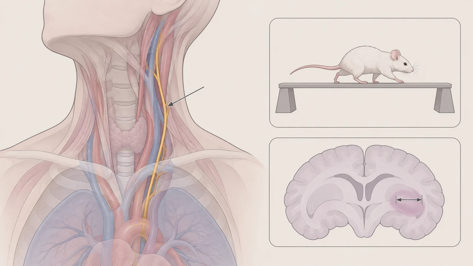
The findings come from a 2022 systematic review and meta-analysis of nine animal studies. Vagus nerve stimulation, or VNS, uses electrical pulses to stimulate the vagus nerve, which can be reached in the neck. The review examined whether it helped cognitive function after traumatic brain injury (TBI), but the clearest pooled results concerned movement and tissue changes—not everyday thinking.
Movement improved more clearly than thinking
Across the review, behavioral testing appeared in five of the nine studies. Two studies reported better performance with VNS on a beam-walking test and a locomotor-placement test. In beam walking, an animal crosses a narrow elevated beam; the placement test records misplaced steps as it moves over a grid. These tests show changes in movement, coordination or limb placement. They do not, by themselves, show that memory, attention or executive function improved.
The review also found that direct cognitive testing was uncommon. One study used a Morris water maze, a test that can assess learning and memory, among a broader group of behavioral measures. That limited coverage makes it difficult to tell whether the movement gains reflected better cognition, improved motor control, or some combination. For readers asking whether stimulation helped an injured animal think more clearly, the review did not have a robust answer.
The distinction matters because movement can improve without demonstrating a change in memory or attention. The positive results are a reason to investigate VNS further after TBI, but they should be described as better performance on particular movement tests rather than restored cognition.
Nine studies used different injuries and stimulation schedules
The review searched six databases for studies published through December 2021. It identified nine eligible experiments involving 36 mice, 268 rats and 27 rabbits. Five studies used a weight-drop injury model, three used fluid percussion, and one used controlled cortical impact. These are different ways of producing experimental brain injury, so the animals did not all represent the same injury conditions.
The stimulation also varied. Most studies applied it within 24 hours of injury, while one began three to four weeks later and two did not specify timing. Five studies used a single 10-to-20-minute session; four used longer schedules ranging from 48 hours to five weeks. Reported frequencies ranged from 5 to 30 hertz, and the stimulation current varied as well. Most animal experiments used implanted electrodes at a cervical branch of the vagus nerve.
Pooling results can help show whether findings point in a similar direction across small experiments. It cannot identify one schedule as the treatment to use: the studies differed in injury models, timing, duration and measured outcomes, and the review did not determine an optimal combination of stimulation settings. The largely implanted animal approach also does not establish that a noninvasive method would produce the same effects.
Less swelling is not the same as less structural damage
Three studies contributed to the pooled analysis of brain water content. The review reported significantly less brain edema—excess fluid and swelling in brain tissue—in animals receiving VNS than in comparison animals. The estimated standardized mean difference was −1.58, with a 95% confidence interval from −2.85 to −0.31. The result supports an effect on this measured swelling outcome in those experiments.
A separate analysis considered lesion size, a measure of structural damage. It included only two studies, and the review concluded that VNS did not significantly reduce lesion size. This result deserves caution: the evidence base was very small, and the two studies differed substantially in their results. Reduced swelling and a smaller lesion are distinct outcomes, so the edema finding should not be taken as proof that stimulation prevented or reversed the underlying tissue damage.
Several biological changes offer clues, not a proven route
The experiments also looked for biological clues. Three studies found lower brain levels of TNF-α, an inflammatory signal, after VNS. One mouse study found less leakage from blood vessels and a smaller rise in AQP-4, a protein that helps move water through brain tissue. A separate study found lower levels of a marker of oxidative damage and higher levels of antioxidant defenses.
These findings fit a possible chain in which inflammation and changes in the barrier around brain blood vessels affect fluid balance and secondary tissue stress after injury. They remain observations from different experiments, however. They do not show which change, if any, caused better movement, or establish a pathway from stimulation to improved memory or attention.
Study quality and testing leave important gaps
The review’s detailed assessment judged the nine experiments to be of low overall quality, with scores from 30% to 60%. None adequately reported a random group-assignment sequence or how assignments were concealed. All had a high risk of performance bias because blinding during treatment was not described. In five studies, the people assessing outcomes were kept unaware of which group the animals were in.
Another design choice affects how the results should be read: when a study measured an outcome more than once, the reviewers selected the time point showing the greatest benefit. That choice could make the pooled estimate look more favorable than a time point selected before the results were known. With few studies and different outcomes, the review also could not reliably assess whether studies with less favorable results were missing from the published record.
Together, sparse cognition-focused tests and weaknesses in allocation and blinding limit confidence about what the movement results mean. Better experiments would need enough animals, planned measurements, and careful methods to distinguish memory or attention from motor ability. The review’s evidence does not establish a treatment effect in people recovering from TBI, and it does not show that noninvasive stimulation would match the predominantly implanted animal interventions.
What this could change
For someone recovering from a head injury, a future stimulation option could be assessed alongside rehabilitation for whether it helps with practical problems such as attention or memory. The animal findings provide a reason to investigate that possibility: movement tests improved in two studies, and a small pooled analysis found less swelling. Turning those observations into a dependable cognitive benefit would require stronger evidence about what stimulation changes, a safe and workable way to deliver it, and human testing that measures cognition directly.
A nearer milestone is replicated animal research with randomized group assignment and blinded assessment, using planned stimulation schedules and tests that separate memory or attention from movement. If that work supports a clear cognitive effect, it could provide a better basis for testing whether stimulation adds a useful option to care after TBI. Until then, the review points to a research question, not an established treatment for cognitive recovery.
Key Sources
- Primary source: Effects and neuroprotective mechanisms of vagus nerve stimulation on cognitive impairment with traumatic brain injury in animal studies: A systematic review and meta-analysis.
DOI or Journal Page · Review or Meta-Analysis · 2022-09-27
Keep exploring