
Research Signal – Source date 2021-03-12
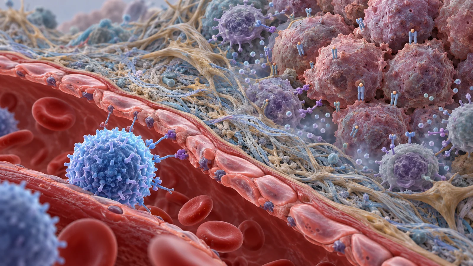
T cells can recognize and kill cancer cells, yet a tumor may still grow. A review published on March 12, 2021, explains why: an effective T-cell response depends on cancer-fighting cells being activated, reaching the tumor, and recognizing and killing their targets there. A tumor can interfere at any of those points. Even when one obstacle is bypassed, another may remain.
The review brings together findings from mouse models, clinical observations, and treatment experience to examine this problem. Its central lesson is that a tumor’s visibility to immune cells and the immune cells’ ability to get inside and function are related, but separate, challenges.
A tumor can change what T cells see
Most cells display small fragments of their internal proteins on their surface, held out by molecules called major histocompatibility complex class I, or MHC I. These fragments offer a sample of what is happening inside the cell. A cytotoxic T cell can inspect them using its T-cell receptor. When a displayed fragment marks a cell as abnormal, the T cell may become activated and kill it.
Cancer cells can make that recognition harder by changing the machinery that processes and displays these fragments. Some tumors reduce MHC I itself; others alter the proteins that cut internal material into fragments or load them onto MHC I. The result can be a changed or diminished set of targets for T cells to inspect.
The kind of defect matters because the review distinguishes between changes that may be reversible and changes caused by genetic damage. In some tumors, gene activity is suppressed without the underlying genes being lost. These “soft” defects may respond to treatments that alter gene regulation, including DNA methyltransferase or histone deacetylation inhibitors. The review also describes combinations involving interferon signaling as a possible way to increase MHC I display. These approaches aim to restore a signal the tumor cell can still produce.
Other tumors have “hard” defects, such as genetic loss affecting MHC I or related components. The review cites estimates that such genetic aberrations occur in 30–40% of human cancers and links them with poor response to cytokine treatment and acquired resistance to anti-PD-1 checkpoint inhibitors. Changing gene activity cannot necessarily repair a gene that is missing or structurally damaged. Gene-delivery experiments using components such as β2-microglobulin or TAP restored MHC I display and T-cell recognition in some models, but delivering those genes to tumors inside the body presents a practical challenge.
The distinction is useful because a strategy that restores suppressed display may not work when the display machinery is genetically broken. Nor does restoring visibility guarantee that enough T cells will reach the tumor or remain active once they arrive.
Engineered cells can bypass one form of hiding
CAR T cells are T cells engineered with a receptor that recognizes a protein on a target cell’s surface. Unlike conventional T-cell recognition, this route does not depend on a tumor presenting a peptide through MHC I. It offers a way around one common visibility problem.
The review describes strong complete-response rates in clinical work on CAR T-cell therapies for blood cancers, particularly those targeting CD19 on B cells. But a tumor can change the target instead. Relapses have occurred when CD19-negative leukemia cells emerge: cells that no longer display the marker targeted by the engineered T cells. A powerful response against cells carrying one marker can therefore leave a route open for cells that lack it.
Researchers tested whether recognizing two markers could narrow that route. In one preclinical leukemia experiment, investigators mixed a small population of cells lacking CD19 or CD22 into a much larger population carrying both markers. The mixture was designed to test whether a rare target-loss population could survive a single-target attack. In mouse models, a CAR designed to recognize both CD19 and CD22 cleared one target-loss leukemia population and significantly inhibited the growth of another. Related tests in patient-derived tumor models found that a CD19-resistant leukemia was cleared by the bivalent CAR, while a leukemia carrying both targets was cleared by either CAR design.
These experiments show the logic of a dual-target approach in the models tested: if a cancer cell loses one marker, the engineered T cell may still recognize the other. They are preclinical findings, not evidence that dual-target CAR T cells reduce relapse or extend survival in patients. Even a CAR that recognizes its targets may have difficulty reaching a solid tumor or functioning in its suppressive environment.
Getting inside depends on signals and access
T cells must travel from the blood into tumor tissue. Chemical signals help guide that movement. The review describes interferon-gamma as one source of signals that can encourage immune cells to enter tumors. It can prompt production of chemokines such as CXCL9 and CXCL10, which attract T cells carrying the matching receptor CXCR3. Studies discussed in the review link these signals and receptors with T-cell recruitment in melanoma; clinical observations also associate higher tumor levels of these chemokines and T-cell receptors with greater infiltration and, in some settings, more favorable outcomes.
Tumor blood vessels and tissue structure can make the journey harder. Tumor vessels may be leaky, with uneven blood flow and high pressure in the surrounding tissue. Endothelial cells lining the vessels can also express signals that trap T cells, while factors associated with tumor blood-vessel growth may reduce the adhesion molecules immune cells need to leave the bloodstream and enter tissue.
A tumor containing T cells is therefore informative: it shows that immune cells have reached at least part of the tumor. It does not by itself show that those cells can recognize the cancer, remain active, or eliminate it. Across several cancers, more tumor-infiltrating CD8 T cells have been associated with better survival, but an association does not establish that infiltration alone caused tumor control. The mix of cells matters too; the review contrasts effector T cells and some macrophages associated with favorable outcomes with regulatory T cells and other macrophages that may support tumor growth.
Releasing immune brakes can bring harm
Even after a T cell enters a tumor, local immune-suppressing signals can limit its activity. Inhibitory receptors such as PD-1 and CTLA-4 help regulate T-cell responses; others discussed in the review include LAG-3, TIM-3, and TIGIT. Treatments that block checkpoint signals can restore aspects of tumor-directed immunity, and the review describes checkpoint inhibition as an active area of basic and clinical research.
The same restraint that tumors can exploit also helps keep immune responses from damaging healthy tissue. Releasing inhibitory pathways can activate immune cells beyond the tumor, bringing the risk of immune-related adverse effects. The review notes events including diarrhea, colitis, myocarditis, and endocrine disorders. The therapeutic aim is thus not simply to turn up T-cell activity everywhere: it is to strengthen tumor-specific attack while limiting injury to the body’s own tissues.
Mouse models answer different questions
Mouse experiments help researchers test immune responses in a living system, but different model designs capture different parts of cancer. In syngeneic models, mouse tumor cells are transplanted into mice with intact immune systems. These tumors often grow quickly, making experiments relatively fast and allowing researchers to study immune responses without first transferring additional immune cells. The short growth window can also limit the time available to observe treatment effects, and tumors derived from established cell lines may be more uniform than human cancers. They are often grown under the skin, which may not reproduce the conditions of tumors in their usual organs.
Human-tumor xenografts address a different question by growing human tumor cells in mice. Patient-derived xenografts can preserve features of a patient’s tumor and help assess treatments such as CAR T cells. But the mice must be immunocompromised to prevent their own immune systems from attacking the human cells. Human immune cells can be added in some designs, though it remains uncertain whether a reconstituted immune system behaves as it does in people. A model that includes a human tumor is not automatically a model of the full human immune environment.
The dual-target CAR results sit within this preclinical landscape. They provide proof of concept in engineered mouse experiments that recognizing two markers can constrain particular escape populations. They do not settle how the approach will perform across the diversity of human tumors, their blood vessels, and their local immune suppression. The models are useful precisely when their strengths and limits are kept in view.
What longer-lasting T-cell treatment could change
If future approaches can preserve tumor-specific recognition while improving T-cell entry and persistence, they could make immune therapies more capable of reaching and attacking cancers that currently evade one part of the response. That payoff depends on solving several problems together: a tumor may alter what it displays, obstruct immune-cell access, and suppress cells that arrive. The review points toward a multifaceted direction for research, not a single remedy already shown to overcome every defense.
Key Sources
- Primary source: Investigating T Cell Immunity in Cancer: Achievements and Prospects.
DOI or Journal Page · Review or Meta-Analysis · 2021-03-12
Keep exploring