
Research Signal – Source date 2021-01-20
A PD-L1 result can depend on which cells carry the protein. In studies reviewed for a 2021 overview of lung cancer immunotherapy, higher PD-L1 on macrophages—immune cells found in tumors—was associated with longer survival among some patients receiving immunotherapy. The same pattern was not seen when PD-L1 was measured on cancer cells or among patients who had not received immunotherapy.
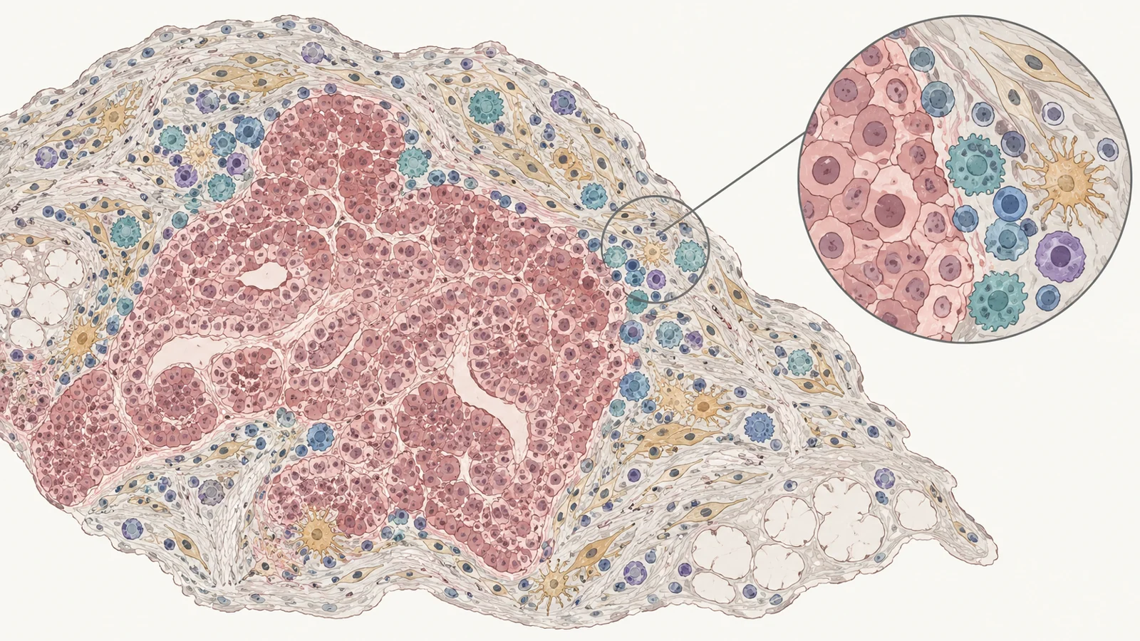
The review did not test a new treatment or a new decision rule. It brought together earlier tissue studies and comparisons of testing methods. Its central technical idea is to examine multiple markers on one tissue slice while preserving information about which cells carry them and where they sit. That can offer a richer view of a biopsy, but the findings were still research evidence requiring broader clinical validation.
A biopsy has limited room for separate stains
A pathologist may use immunohistochemistry or immunofluorescence to detect proteins in a tissue sample. Conventional versions generally stain one to three targets on a preserved slice. Testing more proteins may require additional slices, which can be difficult when a biopsy contains only a small amount of tissue beyond what is needed to identify the cancer.
Separate slices also make it harder to tell whether two proteins occur in the same cell, in neighboring cells, or in different parts of the tumor. Those relationships may matter when the question is not just whether a marker is present, but which cell carries it and where that cell is located.
Multiplex immunohistochemistry or immunofluorescence, abbreviated mIHC/IF, aims to keep those details together. The tissue is stained and imaged in repeated rounds on the same slice. Between rounds, antibodies are removed; depending on the method, earlier color signals are either cleared or preserved for later image analysis. Images are then combined and analyzed so researchers can identify cell types, count them, and examine their positions relative to tumor tissue and other cells.
The review describes several approaches, including methods that strip antibodies between staining rounds and fluorescent methods that preserve each signal while removing antibodies. Some imaging systems can detect many markers on one slice. The practical point is that multiple measurements can be linked back to the same tissue region, rather than inferred across separate slices.
Location changed the survival association
Where immune cells sit can distinguish one tissue finding from another. In a study of 112 people with non-small-cell lung cancer, researchers examined tissue from 61 patients who had received chemotherapy before surgery and 51 who had not. Among the chemotherapy group, a higher density of CD4-positive T cells within the tumor epithelium—the layer containing the cancer cells—was associated with longer overall survival. A higher density in the surrounding stroma, the supportive tissue around the tumor, was not associated with survival in that group.
Other studies summarized in the review also linked spatial patterns with outcomes. For example, one reported that more regulatory T cells in the tumor core were associated with worse survival, while the presence of nearby CD8-positive T cells modified that association. Such findings suggest that cell counts alone may miss meaningful differences in tissue organization. They do not establish that moving or changing those cells would alter a patient’s outcome.
The cell carrying PD-L1 mattered in treated groups
PD-L1 is a protein measured in immunotherapy testing. The review describes studies in which its association with survival varied by the cell type carrying it and by whether patients received immune checkpoint treatment.
In one study, tissue from 69 patients with non-small-cell lung cancer who received immunotherapy was compared with tissue from 258 who did not. Among treated patients, higher levels of both PD-L1 and a second protein, CMTM6, on macrophages identified by the CD68 marker were associated with longer overall survival (hazard ratio 0.38, 95% confidence interval 0.16–0.92). The association was not significant when the same proteins were assessed in tumor cells, and the macrophage pattern was not observed in the untreated group.
A separate study examined 62 patients treated with pembrolizumab, nivolumab, or atezolizumab and 425 who had not received immunotherapy. Among the treated patients, higher PD-L1 in macrophages was associated with longer overall survival. The review reports no such association for PD-L1 measured in tumor cells or stromal cells, or for macrophage PD-L1 in the untreated group. These are associations from prior studies, not proof that macrophage PD-L1 predicts benefit for an individual patient or should guide treatment on its own.
A promising comparison spans many cancer types
The review also cites an analysis comparing biomarker approaches to predicting response to PD-1 or PD-L1 inhibitors. Across 24 studies, more than 8,000 patients, and over ten types of solid tumor, multiplex testing had a higher reported area under the curve than PD-L1 immunohistochemistry, gene-expression profiles, or tumor mutation burden. The area under the curve measures how well a test separates people with different outcomes in the analyzed data. The reported value for multiplex testing was 0.79, compared with 0.65 for PD-L1 immunohistochemistry, 0.65 for gene-expression profiles, and 0.69 for tumor mutation burden.
That comparison pooled varied cancers and studies. It is not a validated performance estimate for every lung cancer type, and it does not show that using a multiplex result improves treatment outcomes. Lung cancer findings in the same review point to why broad generalization is difficult: among 536 people with non-small-cell lung cancer, a higher density of tumor-associated neutrophils was associated with better prognosis in squamous cell carcinoma but worse prognosis in adenocarcinoma. A cell count or marker pattern may carry different associations across tumor subtypes.
More colors bring laboratory tradeoffs
Multiplex testing adds technical and practical demands. Antibodies can react in unwanted ways, and fluorescent signals can overlap. Some methods reduce tissue damage by removing antibodies while preserving the detected signals, but more colors require specialized imaging equipment and analysis software to separate those signals reliably. The review notes that equipment, computing, storage, and analysis capacity can limit wider use.
The image analysis itself also involves choices about which regions of a tissue slice to examine. If only selected areas are measured, a result may not represent the whole tumor. A rare cell group or a different region could be missed, especially in a tissue sample with variation from place to place. Whole-slice analysis was still being evaluated in the review’s account.
What richer biopsy maps could change for lung cancer care
If larger studies confirm these patterns and laboratories can make the methods reliable and accessible, a biopsy map could help clinicians interpret immune markers in the context of the cells and tumor regions that carry them. That could eventually support more informative treatment research and patient selection than a single stain alone. At the time of the 2021 review, however, the approach still needed rigorous validation before routine clinical use.
Key Sources
- Primary source: [Overview of Multiplex Immunohistochemistry and Immunofluorescence Techniques in the Lung Cancer Immunotherapy].
DOI or Journal Page · Review or Meta-Analysis · 2021-01-20
Keep exploring